 Movie
Movie Controller

[English] 日本語
 Yorodumi
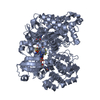
- PDB-9pxt: Cryo-EM structure of NapA, the periplasmic nitrate reductase from... -
+ Open data

- Basic information
| Entry | Database: PDB / ID: 9pxt | ||||||||||||||||||||||||
|---|---|---|---|---|---|---|---|---|---|---|---|---|---|---|---|---|---|---|---|---|---|---|---|---|---|
| Title | Cryo-EM structure of NapA, the periplasmic nitrate reductase from Campylobacter jejuni | ||||||||||||||||||||||||
 Components Components | Periplasmic nitrate reductase | ||||||||||||||||||||||||
 Keywords Keywords | OXIDOREDUCTASE / Periplasmic nitrate reductase / molybdenum enzymes / cryo-EM / MEMBRANE PROTEIN / METAL BINDING PROTEIN | ||||||||||||||||||||||||
| Function / homology |  Function and homology information Function and homology informationnitrate reductase (cytochrome) / nitrate reductase (cytochrome) activity / nitrate reductase complex / Mo-molybdopterin cofactor biosynthetic process / molybdenum ion binding / molybdopterin cofactor binding / cellular respiration / nitrate assimilation / 4 iron, 4 sulfur cluster binding / periplasmic space ...nitrate reductase (cytochrome) / nitrate reductase (cytochrome) activity / nitrate reductase complex / Mo-molybdopterin cofactor biosynthetic process / molybdenum ion binding / molybdopterin cofactor binding / cellular respiration / nitrate assimilation / 4 iron, 4 sulfur cluster binding / periplasmic space / electron transfer activity / iron ion binding / membrane Similarity search - Function | ||||||||||||||||||||||||
| Biological species |   Campylobacter jejuni (Campylobacter) Campylobacter jejuni (Campylobacter) | ||||||||||||||||||||||||
| Method | ELECTRON MICROSCOPY / single particle reconstruction / cryo EM / Resolution: 3 Å | ||||||||||||||||||||||||
 Authors Authors | Thach, T. / Subramanian, R. | ||||||||||||||||||||||||
| Funding support | 1items
| ||||||||||||||||||||||||
 Citation Citation | Journal: J Biol Chem / Year: 2025 Title: Structure and substrate promiscuity of Campylobacter jejuni periplasmic nitrate reductase (Nap) and phylogenetic analysis of Nap homologs. Authors: Nitai C Giri / Trung Thach / KanagaVijayan Dhanabalan / Mintare Cesiunaite / Manohar Radhakrishnan / Lahiru Wedasingha / Nicholas Manicke / Michael Wells / Maciej Szaleniec / Ramaswamy ...Authors: Nitai C Giri / Trung Thach / KanagaVijayan Dhanabalan / Mintare Cesiunaite / Manohar Radhakrishnan / Lahiru Wedasingha / Nicholas Manicke / Michael Wells / Maciej Szaleniec / Ramaswamy Subramanian / Partha Basu /   Abstract: Periplasmic nitrate reductase NapA is a member of the DMSO reductase (DMSOR) superfamily, which catalyzes the reduction of nitrate to nitrite. Campylobacter jejuni NapA (CjNapA) is notably larger ...Periplasmic nitrate reductase NapA is a member of the DMSO reductase (DMSOR) superfamily, which catalyzes the reduction of nitrate to nitrite. Campylobacter jejuni NapA (CjNapA) is notably larger compared to other structurally characterized NapA. Herein, we present the cryo-EM structure of CjNapA, the first of its kind from any ε-proteobacteria, revealing three lysine-rich insertions that could affect the substrate channel, potentially enhancing the affinity towards nitrate and other anionic substrates. Here, we report that wild-type CjNapA and NapA-C176D variants can reduce chlorate, perchlorate, and nitrate. However, the perchlorate and chlorate reductions by the CjNapA C176D variant are considerably slower, even though the perchlorate reductase has an Asp coordination to Mo. Molecular Dynamics (MD) simulations were performed to investigate the impact of the C176D mutation on substrate affinity and protein flexibility. Structural and kinetic comparisons with perchlorate reductase support evolutionary tuning for a desired function. Finally, structural comparisons with other structurally characterized NapAs also suggest the role of proximal pterin in CjNapA in electron transfer to the Mo center. | ||||||||||||||||||||||||
| History |
|
- Structure visualization
| Structure viewer | Molecule: MolmilJmol/JSmol |
|---|
- Downloads & links
-Download
| PDBx/mmCIF format | 9pxt.cif.gz | 228.6 KB | Display | PDBx/mmCIF format |
|---|---|---|---|---|
| PDB format | pdb9pxt.ent.gz | 143.5 KB | Display | PDB format |
| PDBx/mmJSON format | 9pxt.json.gz | Tree view | PDBx/mmJSON format | |
| Others |  Other downloads Other downloads |
-Validation report
| Arichive directory | https://data.pdbj.org/pub/pdb/validation_reports/px/9pxtftp://data.pdbj.org/pub/pdb/validation_reports/px/9pxt | HTTPS FTP |
|---|
-Related structure data
| Related structure data |  71994MC M: map data used to model this data C: citing same article ( |
|---|---|
| Similar structure data |
-Links
 PDBj
PDBj


- Assembly
| Deposited unit | 
|
|---|---|
| 1 |
|
-Components
| #1: Protein | Mass: 105092.828 Da / Num. of mol.: 1 Source method: isolated from a genetically manipulated source Source: (gene. exp.) Campylobacter jejuni (Campylobacter) / Gene: napA, Cj0780 / Production host: | ||||
|---|---|---|---|---|---|
| #2: Chemical | ChemComp-SF4 /   Mass: 351.640 Da / Num. of mol.: 1 / Source method: obtained synthetically / Formula: Fe4S4 / Feature type: SUBJECT OF INVESTIGATION Mass: 351.640 Da / Num. of mol.: 1 / Source method: obtained synthetically / Formula: Fe4S4 / Feature type: SUBJECT OF INVESTIGATION | ||||
| #3: Chemical | ChemComp-MO / 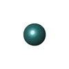  Mass: 95.940 Da / Num. of mol.: 1 / Source method: obtained synthetically / Formula: Mo / Feature type: SUBJECT OF INVESTIGATION Mass: 95.940 Da / Num. of mol.: 1 / Source method: obtained synthetically / Formula: Mo / Feature type: SUBJECT OF INVESTIGATION | ||||
| #4: Chemical | 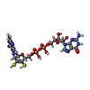  Mass: 740.557 Da / Num. of mol.: 2 / Source method: obtained synthetically / Formula: C20H26N10O13P2S2 / Feature type: SUBJECT OF INVESTIGATION Mass: 740.557 Da / Num. of mol.: 2 / Source method: obtained synthetically / Formula: C20H26N10O13P2S2 / Feature type: SUBJECT OF INVESTIGATIONHas ligand of interest | Y | Has protein modification | N | |
-Experimental details
-Experiment
| Experiment | Method: ELECTRON MICROSCOPY |
|---|---|
| EM experiment | Aggregation state: PARTICLE / 3D reconstruction method: single particle reconstruction |
- Sample preparation
| Component | Name: Structure of NapA:Moco[4Fe4S] / Type: COMPLEX / Entity ID: #1 / Source: RECOMBINANT | |||||||||||||||||||||||||
|---|---|---|---|---|---|---|---|---|---|---|---|---|---|---|---|---|---|---|---|---|---|---|---|---|---|---|
| Molecular weight | Value: 0.084 MDa / Experimental value: YES | |||||||||||||||||||||||||
| Source (natural) | Organism: Campylobacter jejuni (Campylobacter) | |||||||||||||||||||||||||
| Source (recombinant) | Organism: | |||||||||||||||||||||||||
| Buffer solution | pH: 7.5 Details: 25 mM Hepes 7.0, 100 mM NaCl, 0.5 mM TCEP, 2% glycerol | |||||||||||||||||||||||||
| Buffer component |
| |||||||||||||||||||||||||
| Specimen | Conc.: 1 mg/ml / Embedding applied: NO / Shadowing applied: NO / Staining applied: NO / Vitrification applied: YES | |||||||||||||||||||||||||
| Specimen support | Grid type: UltrAuFoil R1.2/1.3 | |||||||||||||||||||||||||
| Vitrification | Instrument: FEI VITROBOT MARK IV / Cryogen name: ETHANE / Humidity: 100 % / Chamber temperature: 277.15 K / Details: vitrification |
- Electron microscopy imaging
| Experimental equipment |  Model: Titan Krios / Image courtesy: FEI Company |
|---|---|
| Microscopy | Model: TFS KRIOS |
| Electron gun | Electron source:  FIELD EMISSION GUN / Accelerating voltage: 300 kV / Illumination mode: FLOOD BEAM FIELD EMISSION GUN / Accelerating voltage: 300 kV / Illumination mode: FLOOD BEAM |
| Electron lens | Mode: BRIGHT FIELD / Nominal defocus max: 2000 nm / Nominal defocus min: 700 nm / Cs: 0.1 mm / C2 aperture diameter: 70 µm |
| Specimen holder | Cryogen: NITROGEN |
| Image recording | Average exposure time: 1.8 sec. / Electron dose: 56.8 e/Å2 / Film or detector model: GATAN K3 BIOCONTINUUM (6k x 4k) / Num. of grids imaged: 1 / Num. of real images: 60 |
| EM imaging optics | Energyfilter name: GIF Bioquantum |
- Processing
| EM software |
| ||||||||||||||||||||||||
|---|---|---|---|---|---|---|---|---|---|---|---|---|---|---|---|---|---|---|---|---|---|---|---|---|---|
| CTF correction | Type: PHASE FLIPPING AND AMPLITUDE CORRECTION | ||||||||||||||||||||||||
| Particle selection | Num. of particles selected: 200982 | ||||||||||||||||||||||||
| 3D reconstruction | Resolution: 3 Å / Resolution method: FSC 0.143 CUT-OFF / Num. of particles: 131448 / Symmetry type: POINT | ||||||||||||||||||||||||
| Atomic model building | Protocol: AB INITIO MODEL / Space: REAL / Details: real refinement was done using Phenix | ||||||||||||||||||||||||
| Atomic model building | Chain residue range: 1-924 / Details: The initial model consisted of monomer / Source name: AlphaFold / Type: in silico model | ||||||||||||||||||||||||
| Refinement | Cross valid method: NONE Stereochemistry target values: GeoStd + Monomer Library + CDL v1.2 | ||||||||||||||||||||||||
| Displacement parameters | Biso mean: 34.67 Å2 | ||||||||||||||||||||||||
| Refine LS restraints |
|
