Movie
Movie Controller

[English] 日本語
 Yorodumi
- PDB-9pse: In situ MicroED structure of IL-5 activated human eosinophil majo... -
+ Open data

- Basic information
| Entry | Database: PDB / ID: 9pse | ||||||||||||||||||
|---|---|---|---|---|---|---|---|---|---|---|---|---|---|---|---|---|---|---|---|
| Title | In situ MicroED structure of IL-5 activated human eosinophil major basic protein-1 | ||||||||||||||||||
 Components Components | Bone marrow proteoglycan | ||||||||||||||||||
 Keywords Keywords | IMMUNE SYSTEM / Effector / Nanocrystal / In-situ / Intracellular | ||||||||||||||||||
| Function / homology |  Function and homology information Function and homology informationextracellular matrix structural constituent conferring compression resistance / transport vesicle / heparin binding / carbohydrate binding / extracellular matrix / ficolin-1-rich granule lumen / defense response to bacterium / immune response / Neutrophil degranulation / extracellular exosome / extracellular region Similarity search - Function | ||||||||||||||||||
| Biological species |  Homo sapiens (human) Homo sapiens (human) | ||||||||||||||||||
| Method | ELECTRON CRYSTALLOGRAPHY / electron crystallography / cryo EM / Resolution: 3.18 Å | ||||||||||||||||||
 Authors Authors | Yang, J.E. / Bingman, C.A. / Mitchell, J. / Mosher, D. / Wright, E.R. | ||||||||||||||||||
| Funding support |  United States, 5items United States, 5items
| ||||||||||||||||||
 Citation Citation | Journal: To Be Published Title: In situ microED structure of the Eosinophil major basic protein-1 Authors: Yang, J.E. / Bingman, C.A. / Mitchell, J. / Mosher, D. / Wright, E.R. | ||||||||||||||||||
| History |
|
- Structure visualization
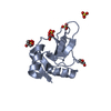
| Structure viewer | Molecule: MolmilJmol/JSmol |
|---|
- Downloads & links
-Download
| PDBx/mmCIF format | 9pse.cif.gz | 119.7 KB | Display | PDBx/mmCIF format |
|---|---|---|---|---|
| PDB format | pdb9pse.ent.gz | 73.9 KB | Display | PDB format |
| PDBx/mmJSON format | 9pse.json.gz | Tree view | PDBx/mmJSON format | |
| Others |  Other downloads Other downloads |
-Validation report
| Arichive directory | https://data.pdbj.org/pub/pdb/validation_reports/ps/9pseftp://data.pdbj.org/pub/pdb/validation_reports/ps/9pse | HTTPS FTP |
|---|
-Related structure data
| Related structure data | |
|---|---|
| Similar structure data |


-Links
 PDBj
PDBj


- Assembly
| Deposited unit | 
| ||||||||||||
|---|---|---|---|---|---|---|---|---|---|---|---|---|---|
| 1 | 
| ||||||||||||
| 2 | 
| ||||||||||||
| Unit cell |
|
-Components
| #1: Protein | Mass: 13694.769 Da / Num. of mol.: 2 Source method: isolated from a genetically manipulated source Details: Human MBP-1 derives from a precursor or proform proMBP coded by gene PRG2 in eosinophils. Protein MBP-1 naturally forms intragranular nanocrystals that get disassembled upon activation, including cytokine IL-5. Source: (gene. exp.) Homo sapiens (human) / Gene: PRG2, MBP / Production host: Homo sapiens (human) / References: UniProt: P13727#2: Chemical |   Mass: 35.453 Da / Num. of mol.: 2 / Source method: obtained synthetically / Formula: Cl / Feature type: SUBJECT OF INVESTIGATION Mass: 35.453 Da / Num. of mol.: 2 / Source method: obtained synthetically / Formula: Cl / Feature type: SUBJECT OF INVESTIGATION#3: Water | ChemComp-HOH / |  Mass: 18.015 Da / Num. of mol.: 3 / Source method: isolated from a natural source / Formula: H2O Mass: 18.015 Da / Num. of mol.: 3 / Source method: isolated from a natural source / Formula: H2OHas ligand of interest | Y | Has protein modification | Y | Nonpolymer details | Authors state that the chloride ions A301 and B301 modeled could be bromide ions. | |
|---|
-Experimental details
-Experiment
| Experiment | Method: ELECTRON CRYSTALLOGRAPHY |
|---|---|
| EM experiment | Aggregation state: CELL / 3D reconstruction method: electron crystallography |
- Sample preparation
| Component | Name: In situ MicroED structure of the IL-5 activated human eosinophil major basic protein-1 Type: CELL Details: Mature human eosinophil cells were collected and purified from human donor blood. This was followed by eosinophil resting and deposition on EM grids. The cells were then activated by IL-5 ...Details: Mature human eosinophil cells were collected and purified from human donor blood. This was followed by eosinophil resting and deposition on EM grids. The cells were then activated by IL-5 for 1 hour prior to grid plunge-freezing. Cryo-FIB milling of individual eosinophil cells was then performed to expose unperturbed cytosolic secretory granules, inside which nanocrystals of the major basic protein-1 were located. MicroED data were collected on these crystals for in situ structure determination. Entity ID: #1 / Source: NATURAL |
|---|---|
| Source (natural) | Organism: Homo sapiens (human) / Cellular location: cytoplasm / Organ: blood / Organelle: secretory granule |
| Buffer solution | pH: 7.4 Details: Harvested from human donor peripheral blood and rested in 1640 RPMI medium supplemented with 0.1% human serum albumin (HSA) |
| Specimen | Embedding applied: NO / Shadowing applied: NO / Staining applied: NO / Vitrification applied: YES |
| Specimen support | Details: Pre-vacuum was 1 x 10⁻³ Pa and the process pressure was 1 Pa Grid material: GOLD / Grid mesh size: 200 divisions/in. / Grid type: Quantifoil |
| Vitrification | Instrument: LEICA EM GP / Cryogen name: ETHANE / Humidity: 100 % |
-Data collection
| Experimental equipment |  Model: Titan Krios / Image courtesy: FEI Company | ||||||||||||||||||||||||||||||||
|---|---|---|---|---|---|---|---|---|---|---|---|---|---|---|---|---|---|---|---|---|---|---|---|---|---|---|---|---|---|---|---|---|---|
| Microscopy | Model: TFS KRIOS | ||||||||||||||||||||||||||||||||
| Electron gun | Electron source:  FIELD EMISSION GUN / Accelerating voltage: 300 kV / Illumination mode: FLOOD BEAM FIELD EMISSION GUN / Accelerating voltage: 300 kV / Illumination mode: FLOOD BEAM | ||||||||||||||||||||||||||||||||
| Electron lens | Mode: DIFFRACTION / Nominal defocus max: 0 nm / Nominal defocus min: 0 nm / Cs: 2.7 mm / Alignment procedure: BASIC | ||||||||||||||||||||||||||||||||
| Specimen holder | Cryogen: NITROGEN / Specimen holder model: FEI TITAN KRIOS AUTOGRID HOLDER | ||||||||||||||||||||||||||||||||
| Image recording | Average exposure time: 1 sec. / Electron dose: 0.1 e/Å2 / Film or detector model: FEI CETA (4k x 4k) / Num. of diffraction images: 40 Details: Ten datasets, each with 40 images, were scaled together using XSCALE. The electron dose per image was 0.1 e/A^2. | ||||||||||||||||||||||||||||||||
| EM diffraction shell | Resolution: 3→30.69 Å / Fourier space coverage: 85.7 % / Multiplicity: 4.45 / Num. of structure factors: 6805 / Phase residual: 38.78 ° | ||||||||||||||||||||||||||||||||
| EM diffraction stats | Fourier space coverage: 85.7 % / High resolution: 3 Å / Num. of intensities measured: 35354 / Num. of structure factors: 6805 Phase error rejection criteria: no rejections based on phase error Rmerge: 63.1 | ||||||||||||||||||||||||||||||||
| Reflection | Resolution: 3→30.69 Å / Num. obs: 6805 / % possible obs: 85.7 % / Biso Wilson estimate: 47.9 Å2 / CC1/2: 0.682 / Rmerge(I) obs: 0.66 / Rrim(I) all: 0.724 / Net I/σ(I): 2.47 / Num. measured all: 35354 | ||||||||||||||||||||||||||||||||
| Reflection shell | Diffraction-ID: 1
|
- Processing
| Software |
| ||||||||||||||||||||||||||||
|---|---|---|---|---|---|---|---|---|---|---|---|---|---|---|---|---|---|---|---|---|---|---|---|---|---|---|---|---|---|
| EM software |
| ||||||||||||||||||||||||||||
| EM 3D crystal entity | ∠α: 90 ° / ∠β: 91.2 ° / ∠γ: 90 ° / A: 30.2 Å / B: 57.8 Å / C: 58.6 Å / Space group name: P1211 / Space group num: 3 | ||||||||||||||||||||||||||||
| CTF correction | Type: NONE | ||||||||||||||||||||||||||||
| 3D reconstruction | Resolution: 3.18 Å / Resolution method: DIFFRACTION PATTERN/LAYERLINES / Symmetry type: 3D CRYSTAL | ||||||||||||||||||||||||||||
| Atomic model building | B value: 40 / Protocol: OTHER / Space: RECIPROCAL / Target criteria: Maximum likelihood Details: Iterative refinement using phenix.refine and model building in COOT | ||||||||||||||||||||||||||||
| Atomic model building | 3D fitting-ID: 1 / Chain-ID: A / Chain residue range: 107-222
| ||||||||||||||||||||||||||||
| Refinement | Resolution: 3.18→3.18 Å / SU ML: 0.5783 / Cross valid method: FREE R-VALUE / σ(F): 1.35 / Phase error: 32.2491 Stereochemistry target values: GeoStd + Monomer Library + CDL v1.2
| ||||||||||||||||||||||||||||
| Solvent computation | Shrinkage radii: 0.9 Å / VDW probe radii: 1.1 Å / Solvent model: FLAT BULK SOLVENT MODEL | ||||||||||||||||||||||||||||
| Displacement parameters | Biso mean: 40 Å2 | ||||||||||||||||||||||||||||
| Refine LS restraints |
| ||||||||||||||||||||||||||||
| LS refinement shell |
|