+ Open data
- Basic information
| Entry | Database: PDB / ID: 9peb | ||||||
|---|---|---|---|---|---|---|---|
| Title | Cryo-EM structure of Arabidopsis thaliana Met1 | ||||||
 Components Components | DNA (cytosine-5)-methyltransferase 1 | ||||||
 Keywords Keywords | TRANSFERASE / DNA (cytosine-5)-methyltransferase | ||||||
| Function / homology |  Function and homology information Function and homology informationzygote asymmetric cytokinesis in embryo sac / negative regulation of flower development / DNA-mediated transformation / DNA (cytosine-5-)-methyltransferase / DNA (cytosine-5-)-methyltransferase activity / DNA methylation-dependent constitutive heterochromatin formation / methyltransferase activity / methylation / chromatin binding / DNA binding / nucleus Similarity search - Function | ||||||
| Biological species |  | ||||||
| Method | ELECTRON MICROSCOPY / single particle reconstruction / cryo EM / Resolution: 3.13 Å | ||||||
 Authors Authors | Lu, J. / Chen, X. / Song, J. | ||||||
| Funding support |  United States, 1items United States, 1items
| ||||||
 Citation Citation |  Journal: Plant Cell / Year: 2025 Journal: Plant Cell / Year: 2025Title: Structure and autoinhibitory regulation of MET1 in the maintenance of plant CG methylation. Authors: Jiuwei Lu / Xinyi Chen / Jian Fang / Daniel Li / Huy Le / Xuehua Zhong / Jikui Song /  Abstract: Plant DNA METHYLTRANSFERASE 1 (MET1) is responsible for maintaining genome-wide CG methylation. Its dysregulation has been linked to profound biological disruptions, including genomic instability and ...Plant DNA METHYLTRANSFERASE 1 (MET1) is responsible for maintaining genome-wide CG methylation. Its dysregulation has been linked to profound biological disruptions, including genomic instability and developmental defects. However, the exact mechanism by which MET1 orchestrates these vital functions and coordinates its various domains to shape the plant-specific epigenome remains unknown. Here, we report the cryo-EM structure of Arabidopsis thaliana MET1 (AtMET1), revealing an autoinhibitory mechanism that governs its DNA methylation activity. Between the two replication-foci-target sequence (RFTS) domains in AtMET1, the second RFTS domain (RFTS2) directly associates with the methyltransferase (MTase) domain, thereby inhibiting substrate-binding activity. Compared to DNMT1, AtMET1 lacks the CXXC domain and its downstream autoinhibitory linker, featuring only limited RFTS2-MTase interactions, resulting in a much-reduced autoinhibitory contact. In line with this difference, the DNA methylation activity of AtMET1 displays less temperature dependence than that of DNMT1, potentially allowing MET1 to maintain its activity across diverse temperature conditions. We further report the structure of AtMET1 bound to hemimethylated CG (hmCG) DNA, unveiling the molecular basis for substrate binding and CG recognition by AtMET1, and an activation mechanism that involves a coordinated conformational shift between two structural elements of its active site. In addition, our combined structural and biochemical analysis highlights distinct functionalities between the two RFTS domains of AtMET1, unraveling their evolutionary divergence from the DNMT1 RFTS domain. Together, this study offers a framework for understanding the structure and mechanism of AtMET1, with profound implications for the maintenance of CG methylation in plants. | ||||||
| History |
|
- Structure visualization
| Structure viewer | Molecule:  Molmil Molmil Jmol/JSmol Jmol/JSmol |
|---|
- Downloads & links
- Download
| PDBx/mmCIF format |  9peb.cif.gz 9peb.cif.gz | 227.4 KB | Display |  PDBx/mmCIF format PDBx/mmCIF format |
|---|---|---|---|---|
| PDB format |  pdb9peb.ent.gz pdb9peb.ent.gz | 169.1 KB | Display |  PDB format PDB format |
| PDBx/mmJSON format |  9peb.json.gz 9peb.json.gz | Tree view |  PDBx/mmJSON format PDBx/mmJSON format | |
| Others |  Other downloads Other downloads |
-Validation report
| Summary document |  9peb_validation.pdf.gz 9peb_validation.pdf.gz | 1.3 MB | Display |  wwPDB validaton report wwPDB validaton report |
|---|---|---|---|---|
| Full document |  9peb_full_validation.pdf.gz 9peb_full_validation.pdf.gz | 1.3 MB | Display | |
| Data in XML |  9peb_validation.xml.gz 9peb_validation.xml.gz | 38.3 KB | Display | |
| Data in CIF |  9peb_validation.cif.gz 9peb_validation.cif.gz | 56.7 KB | Display | |
| Arichive directory |  https://data.pdbj.org/pub/pdb/validation_reports/pe/9peb https://data.pdbj.org/pub/pdb/validation_reports/pe/9peb ftp://data.pdbj.org/pub/pdb/validation_reports/pe/9peb ftp://data.pdbj.org/pub/pdb/validation_reports/pe/9peb | HTTPS FTP |
-Related structure data
| Related structure data |  71556MC  9pecC  9pedC M: map data used to model this data C: citing same article ( |
|---|---|
| Similar structure data | Similarity search - Function & homology  F&H Search F&H Search |
- Links
Links
- Assembly
| Deposited unit | 
|
|---|---|
| 1 |
|
- Components
| #1: Protein | Mass: 169582.422 Da / Num. of mol.: 1 Source method: isolated from a genetically manipulated source Source: (gene. exp.)  Gene: DMT1, ATHIM, DDM2, DMT01, MET1, MET2, At5g49160, K21P3.3 Production host:  References: UniProt: P34881, DNA (cytosine-5-)-methyltransferase |
|---|---|
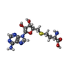
| #2: Chemical | ChemComp-SAH / |
| #3: Chemical | ChemComp-ZN / |
| Has ligand of interest | Y |
| Has protein modification | N |
-Experimental details
-Experiment
| Experiment | Method: ELECTRON MICROSCOPY |
|---|---|
| EM experiment | Aggregation state: PARTICLE / 3D reconstruction method: single particle reconstruction |
- Sample preparation
| Component | Name: Met1 / Type: COMPLEX / Entity ID: #1 / Source: RECOMBINANT |
|---|---|
| Source (natural) | Organism:  |
| Source (recombinant) | Organism:  |
| Buffer solution | pH: 7.5 |
| Specimen | Embedding applied: NO / Shadowing applied: NO / Staining applied: NO / Vitrification applied: YES |
| Vitrification | Cryogen name: ETHANE |
- Electron microscopy imaging
| Experimental equipment |  Model: Titan Krios / Image courtesy: FEI Company |
|---|---|
| Microscopy | Model: TFS KRIOS |
| Electron gun | Electron source:  FIELD EMISSION GUN / Accelerating voltage: 300 kV / Illumination mode: FLOOD BEAM FIELD EMISSION GUN / Accelerating voltage: 300 kV / Illumination mode: FLOOD BEAM |
| Electron lens | Mode: BRIGHT FIELD / Nominal defocus max: 2500 nm / Nominal defocus min: 800 nm |
| Image recording | Electron dose: 50 e/Å2 / Film or detector model: FEI FALCON IV (4k x 4k) |
- Processing
| EM software |
| ||||||||||||||||||||||||
|---|---|---|---|---|---|---|---|---|---|---|---|---|---|---|---|---|---|---|---|---|---|---|---|---|---|
| CTF correction | Type: PHASE FLIPPING AND AMPLITUDE CORRECTION | ||||||||||||||||||||||||
| 3D reconstruction | Resolution: 3.13 Å / Resolution method: FSC 0.143 CUT-OFF / Num. of particles: 172700 / Symmetry type: POINT | ||||||||||||||||||||||||
| Atomic model building | Protocol: AB INITIO MODEL / Space: REAL | ||||||||||||||||||||||||
| Atomic model building | Source name: AlphaFold / Type: in silico model | ||||||||||||||||||||||||
| Refine LS restraints |
|
 Movie
Movie Controller


 PDBj
PDBj


