Movie
Movie Controller


+ Open data

- Basic information
| Entry | Database: PDB / ID: 9pc9 | ||||||||||||||||||||||||||||||
|---|---|---|---|---|---|---|---|---|---|---|---|---|---|---|---|---|---|---|---|---|---|---|---|---|---|---|---|---|---|---|---|
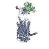
| Title | Structure of Synaptic Vesicle Protein 2A Bound to UCB7361 | ||||||||||||||||||||||||||||||
 Components Components | Synaptic vesicle glycoprotein 2A | ||||||||||||||||||||||||||||||
 Keywords Keywords | MEMBRANE PROTEIN / Synaptic vesicle / SLC22 / Inhibitor | ||||||||||||||||||||||||||||||
| Function / homology |  Function and homology information Function and homology informationToxicity of botulinum toxin type F (botF) / Toxicity of botulinum toxin type D (botD) / Toxicity of botulinum toxin type E (botE) / Toxicity of botulinum toxin type A (botA) / presynaptic active zone / synaptic vesicle priming / transmembrane transporter activity / neuromuscular junction / GABA-ergic synapse / intracellular calcium ion homeostasis ...Toxicity of botulinum toxin type F (botF) / Toxicity of botulinum toxin type D (botD) / Toxicity of botulinum toxin type E (botE) / Toxicity of botulinum toxin type A (botA) / presynaptic active zone / synaptic vesicle priming / transmembrane transporter activity / neuromuscular junction / GABA-ergic synapse / intracellular calcium ion homeostasis / cell-cell junction / synaptic vesicle / synaptic vesicle membrane / neuron projection / protein kinase binding / glutamatergic synapse / endoplasmic reticulum / plasma membrane Similarity search - Function | ||||||||||||||||||||||||||||||
| Biological species |  Homo sapiens (human) Homo sapiens (human) | ||||||||||||||||||||||||||||||
| Method | ELECTRON MICROSCOPY / single particle reconstruction / cryo EM / Resolution: 2.91 Å | ||||||||||||||||||||||||||||||
 Authors Authors | Mittal, A. / Martin, M.F. / Ledecq, M. / Horanyi, P.S. / Coleman, J.A. | ||||||||||||||||||||||||||||||
| Funding support |  United States, 1items United States, 1items
| ||||||||||||||||||||||||||||||
 Citation Citation | Journal: Proc Natl Acad Sci U S A / Year: 2025 Title: Mechanisms underlying allosteric modulation of antiseizure medication binding to synaptic vesicle protein 2A (SV2A). Authors: Anshumali Mittal / Matthew F Martin / Laurent Provins / Adrian Hall / Marie Ledecq / Christian Wolff / Michel Gillard / Peter S Horanyi / Jonathan A Coleman /  Abstract: Brivaracetam (BRV) and levetiracetam (LEV) are antiseizure medications (ASMs); UCB-J is a PET tracer targeting synaptic vesicle protein 2A (SV2A); UCB7361 is closely related to padsevonil, an ...Brivaracetam (BRV) and levetiracetam (LEV) are antiseizure medications (ASMs); UCB-J is a PET tracer targeting synaptic vesicle protein 2A (SV2A); UCB7361 is closely related to padsevonil, an experimental anticonvulsant; while UCB1244283 acts as an allosteric modulator for BRV and LEV binding but not for these other ligands. The SV2A-BRV-UCB1244283 structure reveals how UCB1244283 allosterically enhances BRV binding by occupying an allosteric site near the primary binding site, preventing BRV dissociation. This allosteric site, formed by hydrophobic and uncharged residues, is an uncharacterized small-molecule binding site in SV2A. Structural analysis and mutagenesis demonstrate that an allosteric network between the primary and allosteric sites governs high-affinity ASM binding. Our studies suggest that UCB1244283 selectively binds SV2A over SV2B and SV2C, with specific mutations disrupting binding. Structures of SV2A-UCB-J and SV2A-UCB7361 show that UCB1244283 binding is only possible when the primary site ligand does not overlap with the allosteric site, and that repositioning of Ser601, Thr605, and Leu655 is critical for allosteric ligand binding. Structural comparison of multiple SV2A complexes reveals that primary site occupancy shapes the conformation of the lumenal half of the transmembrane domain, influencing how UCB1244283 binds via a connected network that differentially stabilizes TM1 in either an open or closed conformation and repositions key allosteric and primary site residues. These insights provide a foundation for developing therapeutics targeting the allosteric site and modulating SV2A function. | ||||||||||||||||||||||||||||||
| History |
|
- Structure visualization
| Structure viewer | Molecule: MolmilJmol/JSmol |
|---|
- Downloads & links
-Download
| PDBx/mmCIF format | 9pc9.cif.gz | 172.6 KB | Display | PDBx/mmCIF format |
|---|---|---|---|---|
| PDB format | pdb9pc9.ent.gz | Display | PDB format | |
| PDBx/mmJSON format | 9pc9.json.gz | Tree view | PDBx/mmJSON format | |
| Others |  Other downloads Other downloads |
-Validation report
| Arichive directory | https://data.pdbj.org/pub/pdb/validation_reports/pc/9pc9ftp://data.pdbj.org/pub/pdb/validation_reports/pc/9pc9 | HTTPS FTP |
|---|
-Related structure data
| Related structure data |  71500MC  9ntcC  9pcbC M: map data used to model this data C: citing same article ( |
|---|---|
| Similar structure data |


-Links
 PDBj
PDBj


- Assembly
| Deposited unit | 
|
|---|---|
| 1 |
|
-Components
| #1: Protein | Mass: 82777.031 Da / Num. of mol.: 1 Source method: isolated from a genetically manipulated source Source: (gene. exp.) Homo sapiens (human) / Gene: SV2A, KIAA0736, PSEC0174 / Cell line (production host): tsA201 / Production host: Homo sapiens (human) / References: UniProt: Q7L0J3 |
|---|---|
| #2: Chemical | ChemComp-A1CHN / ( Mass: 430.369 Da / Num. of mol.: 1 / Source method: obtained synthetically / Formula: C15H16F6N4O2S / Feature type: SUBJECT OF INVESTIGATION |
| Has ligand of interest | Y |
| Has protein modification | N |
-Experimental details
-Experiment
| Experiment | Method: ELECTRON MICROSCOPY |
|---|---|
| EM experiment | Aggregation state: PARTICLE / 3D reconstruction method: single particle reconstruction |
- Sample preparation
| Component | Name: SV2A bound to UCB7361 / Type: COMPLEX / Entity ID: #1 / Source: RECOMBINANT |
|---|---|
| Molecular weight | Value: 75.5 kDa/nm / Experimental value: NO |
| Source (natural) | Organism: Homo sapiens (human) |
| Source (recombinant) | Organism: Homo sapiens (human) / Strain: tsa201 |
| Buffer solution | pH: 8 |
| Specimen | Conc.: 7.5 mg/ml / Embedding applied: NO / Shadowing applied: NO / Staining applied: NO / Vitrification applied: YES |
| Specimen support | Grid mesh size: 200 divisions/in. / Grid type: Quantifoil R2/1 |
| Vitrification | Instrument: FEI VITROBOT MARK IV / Cryogen name: ETHANE / Humidity: 100 % / Chamber temperature: 298 K |
- Electron microscopy imaging
| Experimental equipment |  Model: Titan Krios / Image courtesy: FEI Company |
|---|---|
| Microscopy | Model: TFS KRIOS |
| Electron gun | Electron source:  FIELD EMISSION GUN / Accelerating voltage: 300 kV / Illumination mode: FLOOD BEAM FIELD EMISSION GUN / Accelerating voltage: 300 kV / Illumination mode: FLOOD BEAM |
| Electron lens | Mode: BRIGHT FIELD / Nominal magnification: 194000 X / Nominal defocus max: 2000 nm / Nominal defocus min: 500 nm |
| Image recording | Electron dose: 50 e/Å2 / Film or detector model: TFS FALCON 4i (4k x 4k) |
| EM imaging optics | Energyfilter name: TFS Selectris / Energyfilter slit width: 10 eV |
- Processing
| EM software |
| |||||||||||||||||||||||||
|---|---|---|---|---|---|---|---|---|---|---|---|---|---|---|---|---|---|---|---|---|---|---|---|---|---|---|
| CTF correction | Type: PHASE FLIPPING AND AMPLITUDE CORRECTION | |||||||||||||||||||||||||
| 3D reconstruction | Resolution: 2.91 Å / Resolution method: FSC 0.143 CUT-OFF / Num. of particles: 22999 / Symmetry type: POINT | |||||||||||||||||||||||||
| Atomic model building | PDB-ID: 8UO9 Pdb chain-ID: A / Accession code: 8UO9 / Chain residue range: 144-738 / Details: PDB 8UO9 was used for building initial model. / Pdb chain residue range: 144-738 / Source name: PDB / Type: experimental model | |||||||||||||||||||||||||
| Refine LS restraints |
|