[English] 日本語
 Yorodumi
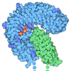
- PDB-9p2r: Extended, CYR715-bound state of Manduca sexta soluble guanylate c... -
+ Open data
- Basic information
| Entry | Database: PDB / ID: 9p2r | |||||||||
|---|---|---|---|---|---|---|---|---|---|---|
| Title | Extended, CYR715-bound state of Manduca sexta soluble guanylate cyclase mutant beta C122S | |||||||||
 Components Components |
| |||||||||
 Keywords Keywords | SIGNALING PROTEIN / Cyclase / NO | |||||||||
| Function / homology |  Function and homology information Function and homology informationguanylate cyclase complex, soluble / guanylate cyclase / guanylate cyclase activity / response to oxygen levels / : / heme binding / GTP binding Similarity search - Function | |||||||||
| Biological species |  Manduca sexta (tobacco hornworm) Manduca sexta (tobacco hornworm) | |||||||||
| Method | ELECTRON MICROSCOPY / single particle reconstruction / cryo EM / Resolution: 3.6 Å | |||||||||
 Authors Authors | Thomas, W.C. / Houghton, K.A. | |||||||||
| Funding support |  United States, 2items United States, 2items
| |||||||||
 Citation Citation |  Journal: Biochemistry / Year: 2025 Journal: Biochemistry / Year: 2025Title: Molecular Aspects of Soluble Guanylate Cyclase Activation and Stimulator Function. Authors: Kimberly A Houghton / William C Thomas / Michael A Marletta /  Abstract: Soluble guanylate cyclases (sGCs) are heme-containing, gas-sensing proteins which catalyze the formation of cGMP from GTP. In humans, sGCs are highly selective sensors of nitric oxide (NO) and play a ...Soluble guanylate cyclases (sGCs) are heme-containing, gas-sensing proteins which catalyze the formation of cGMP from GTP. In humans, sGCs are highly selective sensors of nitric oxide (NO) and play a critical role in NO-based regulation of cardiovascular and pulmonary function. The physiological importance of sGC signaling has led to the development of drugs, known as stimulators and activators, which increase sGC catalytic function. Here we characterize a newly developed stimulator, CYR715, which is a particularly potent stimulator of () sGC catalytic function even in the absence of NO, increasing activity of the NO-free enzyme to 45% of full catalytic activity. CYR715 also increased the catalytic activity of sGC βC122A and βC122S variants, with a marked stimulation of the NO-free βC122S variant to 74% of maximum. High-resolution cryo-electron microscopy structures were solved for CYR715 bound to sGC βC122S revealing that CYR715 occupies the same binding site as the characterized sGC stimulators YC-1 and riociguat. Additionally, the core scaffold of CYR715 makes a binding interaction with βC78 while the flexible tail can interact with αR429 or βY7 and E361. Conformational extension of sGC following NO, YC-1, or CYR715 binding was characterized using small-angle X-ray scattering, revealing that while ligand binding results in sGC extension this extension does not directly correlate to observed activity. This suggests that not all conformational extensions of sGC result in increased catalytic activity, and that effective stimulators assist in converting extension into catalytic function. | |||||||||
| History |
|
- Structure visualization
| Structure viewer | Molecule:  Molmil Molmil Jmol/JSmol Jmol/JSmol |
|---|
- Downloads & links
- Download
| PDBx/mmCIF format |  9p2r.cif.gz 9p2r.cif.gz | 307.7 KB | Display |  PDBx/mmCIF format PDBx/mmCIF format |
|---|---|---|---|---|
| PDB format |  pdb9p2r.ent.gz pdb9p2r.ent.gz | Display |  PDB format PDB format | |
| PDBx/mmJSON format |  9p2r.json.gz 9p2r.json.gz | Tree view |  PDBx/mmJSON format PDBx/mmJSON format | |
| Others |  Other downloads Other downloads |
-Validation report
| Arichive directory |  https://data.pdbj.org/pub/pdb/validation_reports/p2/9p2r https://data.pdbj.org/pub/pdb/validation_reports/p2/9p2r ftp://data.pdbj.org/pub/pdb/validation_reports/p2/9p2r ftp://data.pdbj.org/pub/pdb/validation_reports/p2/9p2r | HTTPS FTP |
|---|
-Related structure data
| Related structure data |  71204MC  9oxnC C: citing same article ( M: map data used to model this data |
|---|---|
| Similar structure data | Similarity search - Function & homology  F&H Search F&H Search |
- Links
Links
- Assembly
| Deposited unit | 
|
|---|---|
| 1 |
|
- Components
| #1: Protein | Mass: 78598.727 Da / Num. of mol.: 1 Source method: isolated from a genetically manipulated source Source: (gene. exp.)  Manduca sexta (tobacco hornworm) Manduca sexta (tobacco hornworm)Production host:  Spodoptera aff. frugiperda 2 RZ-2014 (butterflies/moths) Spodoptera aff. frugiperda 2 RZ-2014 (butterflies/moths)References: UniProt: O77105 |
|---|---|
| #2: Protein | Mass: 68165.930 Da / Num. of mol.: 1 / Mutation: C122S Source method: isolated from a genetically manipulated source Source: (gene. exp.)  Manduca sexta (tobacco hornworm) Manduca sexta (tobacco hornworm)Production host:  Spodoptera aff. frugiperda 2 RZ-2014 (butterflies/moths) Spodoptera aff. frugiperda 2 RZ-2014 (butterflies/moths)References: UniProt: O77106, guanylate cyclase |
| #3: Chemical | ChemComp-G2P / |
| #4: Chemical | ChemComp-HEM / |
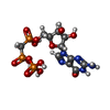
| #5: Chemical | ChemComp-A1CGK / ( Mass: 480.467 Da / Num. of mol.: 1 / Source method: obtained synthetically / Formula: C24H22F2N6O3 / Feature type: SUBJECT OF INVESTIGATION |
| Has ligand of interest | Y |
| Has protein modification | N |
-Experimental details
-Experiment
| Experiment | Method: ELECTRON MICROSCOPY |
|---|---|
| EM experiment | Aggregation state: PARTICLE / 3D reconstruction method: single particle reconstruction |
- Sample preparation
| Component | Name: Ligand-free Manduca sexta soluble guanylase cyclase variant Type: COMPLEX Details: Heterodimeric sGC molecule in the ligand-free, compact state. Beta-C122S mutant variant. Entity ID: #1-#2 / Source: RECOMBINANT | ||||||||||||||||||||||||||||||
|---|---|---|---|---|---|---|---|---|---|---|---|---|---|---|---|---|---|---|---|---|---|---|---|---|---|---|---|---|---|---|---|
| Molecular weight | Value: .147 MDa / Experimental value: NO | ||||||||||||||||||||||||||||||
| Source (natural) | Organism:  Manduca sexta (tobacco hornworm) Manduca sexta (tobacco hornworm) | ||||||||||||||||||||||||||||||
| Source (recombinant) | Organism:  Spodoptera aff. frugiperda 2 RZ-2014 (butterflies/moths) Spodoptera aff. frugiperda 2 RZ-2014 (butterflies/moths) | ||||||||||||||||||||||||||||||
| Buffer solution | pH: 7.5 | ||||||||||||||||||||||||||||||
| Buffer component |
| ||||||||||||||||||||||||||||||
| Specimen | Conc.: 1.5 mg/ml / Embedding applied: NO / Shadowing applied: NO / Staining applied: NO / Vitrification applied: YES Details: Samples were prepared in a Coy anaerobic chamber at RT. Protein was thawed at 4 C, reduced with 10 mM Na2S2O4 for 15 minutes at RT, and desalted using a Zeba spin column equilibrated with ...Details: Samples were prepared in a Coy anaerobic chamber at RT. Protein was thawed at 4 C, reduced with 10 mM Na2S2O4 for 15 minutes at RT, and desalted using a Zeba spin column equilibrated with Buffer, 0.22 um filtered. Protein samples were then diluted to 10 uM in equivalent buffer but with addition of 0.5 mM FOM. Additionally, 250 uM CYR715 (stimulator) and 1 mM GpCpp (non-hydrolyzable substrate-analog) were added to the sample before freezing. | ||||||||||||||||||||||||||||||
| Specimen support | Grid type: Quantifoil R1.2/1.3 | ||||||||||||||||||||||||||||||
| Vitrification | Instrument: FEI VITROBOT MARK IV / Cryogen name: ETHANE / Humidity: 100 % / Chamber temperature: 277 K Details: Cryo-EM samples were prepared by applying 3 ul to a glow-discharged Quantifoil R1.2/1.3 holey-carbon cryo-EM grid. The grid was blotted for 4 s with Whatman #1 filter paper and then plunge- ...Details: Cryo-EM samples were prepared by applying 3 ul to a glow-discharged Quantifoil R1.2/1.3 holey-carbon cryo-EM grid. The grid was blotted for 4 s with Whatman #1 filter paper and then plunge-frozen in liquid ethane with a Mark IV Vitrobot (ThermoFisher) at 4 C and 100% humidity. |
- Electron microscopy imaging
| Experimental equipment |  Model: Titan Krios / Image courtesy: FEI Company |
|---|---|
| Microscopy | Model: TFS KRIOS |
| Electron gun | Electron source:  FIELD EMISSION GUN / Accelerating voltage: 300 kV / Illumination mode: FLOOD BEAM FIELD EMISSION GUN / Accelerating voltage: 300 kV / Illumination mode: FLOOD BEAM |
| Electron lens | Mode: BRIGHT FIELD / Nominal defocus max: 1500 nm / Nominal defocus min: 500 nm / Cs: 2.7 mm / C2 aperture diameter: 100 µm / Alignment procedure: COMA FREE |
| Specimen holder | Specimen holder model: FEI TITAN KRIOS AUTOGRID HOLDER |
| Image recording | Average exposure time: 0.0354 sec. / Electron dose: 1.25 e/Å2 / Film or detector model: GATAN K3 (6k x 4k) / Num. of grids imaged: 1 / Num. of real images: 8433 |
- Processing
| EM software |
| ||||||||||||||||||||||||||||||||||||||||||||
|---|---|---|---|---|---|---|---|---|---|---|---|---|---|---|---|---|---|---|---|---|---|---|---|---|---|---|---|---|---|---|---|---|---|---|---|---|---|---|---|---|---|---|---|---|---|
| CTF correction | Type: PHASE FLIPPING AND AMPLITUDE CORRECTION | ||||||||||||||||||||||||||||||||||||||||||||
| Particle selection | Num. of particles selected: 1226641 Details: Autopicking was used to produce a stack of 2,299,895 particles. Particles showing poor alignment or broken complexes were removed using a series of 2D classification steps, leaving a ...Details: Autopicking was used to produce a stack of 2,299,895 particles. Particles showing poor alignment or broken complexes were removed using a series of 2D classification steps, leaving a particle stack of 902,997 particles that resembled sGC dimers. | ||||||||||||||||||||||||||||||||||||||||||||
| 3D reconstruction | Resolution: 3.6 Å / Resolution method: FSC 0.143 CUT-OFF / Num. of particles: 598571 Details: Non-Uniform (NU) refinement was performed on the higher resolution 3D class. The resulting 3.6 A consensus density resembled previous sGC structures in the extended, NO-bound state. Other ...Details: Non-Uniform (NU) refinement was performed on the higher resolution 3D class. The resulting 3.6 A consensus density resembled previous sGC structures in the extended, NO-bound state. Other refinement strategies were attempted, but did not lead to higher resolution global map densities. To improve local resolution of the domains of CYR715-bound sGC, local refinement was also performed. Separate masks were created for each domain, and local refinement as implemented in cryoSPARC was used to improve the resolution of the catalytic and H-NOX domains of the global map. The subsequent local refinement resulted in 3.5 A and 3.4 A maps for the H-NOX and catalytic domains respectively. These maps were then combined with the consensus map to create the composite map herein. Symmetry type: POINT | ||||||||||||||||||||||||||||||||||||||||||||
| Atomic model building | Protocol: FLEXIBLE FIT / Space: REAL Details: Refinement was performed using iterative rounds of Phenix real space refinement and manual modeling in Coot. Phenix refinement was performed for separate domains of the model using the ...Details: Refinement was performed using iterative rounds of Phenix real space refinement and manual modeling in Coot. Phenix refinement was performed for separate domains of the model using the higher-resolution local maps of those domains. | ||||||||||||||||||||||||||||||||||||||||||||
| Atomic model building | Details: Structure best matched homology and shape with 8HBF. Source name: SwissModel / Type: in silico model |
 Movie
Movie Controller


 PDBj
PDBj


