[English] 日本語
 Yorodumi
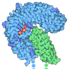
- EMDB-70990: Compact, ligand-free state of Manduca sexta soluble guanylate cyc... -
+ Open data
- Basic information
| Entry |  | |||||||||
|---|---|---|---|---|---|---|---|---|---|---|
| Title | Compact, ligand-free state of Manduca sexta soluble guanylate cyclase mutant beta C122S | |||||||||
 Map data Map data | Composite Map of ligand-free C122S Ms sGC in the compact conformation. A composite map of local refinements of the HNOX and catalytic domains. | |||||||||
 Sample Sample |
| |||||||||
 Keywords Keywords | Cyclase / NO / SIGNALING PROTEIN | |||||||||
| Function / homology |  Function and homology information Function and homology informationguanylate cyclase complex, soluble / guanylate cyclase / guanylate cyclase activity / response to oxygen levels / : / heme binding / GTP binding Similarity search - Function | |||||||||
| Biological species |  Manduca sexta (tobacco hornworm) Manduca sexta (tobacco hornworm) | |||||||||
| Method | single particle reconstruction / cryo EM / Resolution: 3.2 Å | |||||||||
 Authors Authors | Thomas WC / Houghton KA | |||||||||
| Funding support |  United States, 2 items United States, 2 items
| |||||||||
 Citation Citation |  Journal: Biochemistry / Year: 2025 Journal: Biochemistry / Year: 2025Title: Molecular Aspects of Soluble Guanylate Cyclase Activation and Stimulator Function. Authors: Kimberly A Houghton / William C Thomas / Michael A Marletta /  Abstract: Soluble guanylate cyclases (sGCs) are heme-containing, gas-sensing proteins which catalyze the formation of cGMP from GTP. In humans, sGCs are highly selective sensors of nitric oxide (NO) and play a ...Soluble guanylate cyclases (sGCs) are heme-containing, gas-sensing proteins which catalyze the formation of cGMP from GTP. In humans, sGCs are highly selective sensors of nitric oxide (NO) and play a critical role in NO-based regulation of cardiovascular and pulmonary function. The physiological importance of sGC signaling has led to the development of drugs, known as stimulators and activators, which increase sGC catalytic function. Here we characterize a newly developed stimulator, CYR715, which is a particularly potent stimulator of () sGC catalytic function even in the absence of NO, increasing activity of the NO-free enzyme to 45% of full catalytic activity. CYR715 also increased the catalytic activity of sGC βC122A and βC122S variants, with a marked stimulation of the NO-free βC122S variant to 74% of maximum. High-resolution cryo-electron microscopy structures were solved for CYR715 bound to sGC βC122S revealing that CYR715 occupies the same binding site as the characterized sGC stimulators YC-1 and riociguat. Additionally, the core scaffold of CYR715 makes a binding interaction with βC78 while the flexible tail can interact with αR429 or βY7 and E361. Conformational extension of sGC following NO, YC-1, or CYR715 binding was characterized using small-angle X-ray scattering, revealing that while ligand binding results in sGC extension this extension does not directly correlate to observed activity. This suggests that not all conformational extensions of sGC result in increased catalytic activity, and that effective stimulators assist in converting extension into catalytic function. | |||||||||
| History |
|
- Structure visualization
| Supplemental images |
|---|
- Downloads & links
-EMDB archive
| Map data |  emd_70990.map.gz emd_70990.map.gz | 3.8 MB |  EMDB map data format EMDB map data format | |
|---|---|---|---|---|
| Header (meta data) |  emd-70990-v30.xml emd-70990-v30.xml emd-70990.xml emd-70990.xml | 23 KB 23 KB | Display Display |  EMDB header EMDB header |
| Images |  emd_70990.png emd_70990.png | 115.7 KB | ||
| Filedesc metadata |  emd-70990.cif.gz emd-70990.cif.gz | 8.5 KB | ||
| Archive directory |  http://ftp.pdbj.org/pub/emdb/structures/EMD-70990 http://ftp.pdbj.org/pub/emdb/structures/EMD-70990 ftp://ftp.pdbj.org/pub/emdb/structures/EMD-70990 ftp://ftp.pdbj.org/pub/emdb/structures/EMD-70990 | HTTPS FTP |
-Validation report
| Summary document |  emd_70990_validation.pdf.gz emd_70990_validation.pdf.gz | 336.6 KB | Display |  EMDB validaton report EMDB validaton report |
|---|---|---|---|---|
| Full document |  emd_70990_full_validation.pdf.gz emd_70990_full_validation.pdf.gz | 336.1 KB | Display | |
| Data in XML |  emd_70990_validation.xml.gz emd_70990_validation.xml.gz | 6.6 KB | Display | |
| Data in CIF |  emd_70990_validation.cif.gz emd_70990_validation.cif.gz | 7.7 KB | Display | |
| Arichive directory |  https://ftp.pdbj.org/pub/emdb/validation_reports/EMD-70990 https://ftp.pdbj.org/pub/emdb/validation_reports/EMD-70990 ftp://ftp.pdbj.org/pub/emdb/validation_reports/EMD-70990 ftp://ftp.pdbj.org/pub/emdb/validation_reports/EMD-70990 | HTTPS FTP |
-Related structure data
| Related structure data |  9oxnMC  9p2rC C: citing same article ( M: atomic model generated by this map |
|---|---|
| Similar structure data | Similarity search - Function & homology  F&H Search F&H Search |
- Links
Links
| EMDB pages |  EMDB (EBI/PDBe) / EMDB (EBI/PDBe) /  EMDataResource EMDataResource |
|---|---|
| Related items in Molecule of the Month |
- Map
Map
| File |  Download / File: emd_70990.map.gz / Format: CCP4 / Size: 125 MB / Type: IMAGE STORED AS FLOATING POINT NUMBER (4 BYTES) Download / File: emd_70990.map.gz / Format: CCP4 / Size: 125 MB / Type: IMAGE STORED AS FLOATING POINT NUMBER (4 BYTES) | ||||||||||||||||||||||||||||||||||||
|---|---|---|---|---|---|---|---|---|---|---|---|---|---|---|---|---|---|---|---|---|---|---|---|---|---|---|---|---|---|---|---|---|---|---|---|---|---|
| Annotation | Composite Map of ligand-free C122S Ms sGC in the compact conformation. A composite map of local refinements of the HNOX and catalytic domains. | ||||||||||||||||||||||||||||||||||||
| Projections & slices | Image control
Images are generated by Spider. | ||||||||||||||||||||||||||||||||||||
| Voxel size | X=Y=Z: 0.942 Å | ||||||||||||||||||||||||||||||||||||
| Density |
| ||||||||||||||||||||||||||||||||||||
| Symmetry | Space group: 1 | ||||||||||||||||||||||||||||||||||||
| Details | EMDB XML:
|
-Supplemental data
- Sample components
-Entire : Ligand-free Manduca sexta soluble guanylase cyclase variant
| Entire | Name: Ligand-free Manduca sexta soluble guanylase cyclase variant |
|---|---|
| Components |
|
-Supramolecule #1: Ligand-free Manduca sexta soluble guanylase cyclase variant
| Supramolecule | Name: Ligand-free Manduca sexta soluble guanylase cyclase variant type: complex / ID: 1 / Parent: 0 / Macromolecule list: #1-#2 Details: Heterodimeric sGC molecule in the ligand-free, compact state. Beta-C122S mutant variant. |
|---|---|
| Source (natural) | Organism:  Manduca sexta (tobacco hornworm) Manduca sexta (tobacco hornworm) |
| Molecular weight | Theoretical: 147 KDa |
-Macromolecule #1: Soluble guanylyl cyclase alpha-1 subunit
| Macromolecule | Name: Soluble guanylyl cyclase alpha-1 subunit / type: protein_or_peptide / ID: 1 / Number of copies: 1 / Enantiomer: LEVO |
|---|---|
| Source (natural) | Organism:  Manduca sexta (tobacco hornworm) Manduca sexta (tobacco hornworm) |
| Molecular weight | Theoretical: 78.598727 KDa |
| Recombinant expression | Organism:  Spodoptera aff. frugiperda 2 RZ-2014 (butterflies/moths) Spodoptera aff. frugiperda 2 RZ-2014 (butterflies/moths) |
| Sequence | String: MTCPFRRASS QHQFANGGSS APKKPEFRSR TSSVHLTGPE EEDGERNTLT LKHMSEALQL LTAPSNECLH AAVTSLTKNQ SDHYHKYNC LRRLPDDVKT CRNYAYLQEI YDAVRATDSV NTKDFMAKLG EYLILTAFSH NCRLERAFKC LGTNLTEFLT T LDSVHDVL ...String: MTCPFRRASS QHQFANGGSS APKKPEFRSR TSSVHLTGPE EEDGERNTLT LKHMSEALQL LTAPSNECLH AAVTSLTKNQ SDHYHKYNC LRRLPDDVKT CRNYAYLQEI YDAVRATDSV NTKDFMAKLG EYLILTAFSH NCRLERAFKC LGTNLTEFLT T LDSVHDVL HDQDTPLKDE TMEYEANFVC TTSQEGKIQL HLTTESEPVA YLLVGSLKAI AKRLYDTQTD IRLRSYTNDP RR FRYEINA VPLHQKSKED SCELVNEAAS VATSTKVTDL KIGVASFCKA FPWHFITDKR LELVQLGAGF MRLFGTHLAT HGS SLGTYF RLLRPRGVPL DFREILKRVN TPFMFCLKMP GSTALAEGLE IKGQMVFCAE SDSLLFVGSP FLDGLEGLTG RGLF ISDIP LHDATRDVIL VGEQARAQDG LRRRMDKLKN SIEEASKAVD KEREKNVSLL HLIFPPHIAK RLWLGEKIEA KSHDD VTML FSDIVGFTSI CATATPMMVI AMLEDLYSVF DIFCEELDVY KVETIGDAYC VASGLHRKVE THAPQIAWMA LRMVET CAQ HLTHEGNPIK MRIGLHTGTV LAGVVGKTML KYCLFGHNVT LANKFESGSE PLKINVSPTT YEWLIKFPGF DMEPRDR SC LPNSFPKDIH GTCYFLHKYT HPGTDPGEPQ VKHIREALKD YGIGQANSTD VDTEEPT UniProtKB: guanylate cyclase |
-Macromolecule #2: Guanylate cyclase soluble subunit beta-1
| Macromolecule | Name: Guanylate cyclase soluble subunit beta-1 / type: protein_or_peptide / ID: 2 / Number of copies: 1 / Enantiomer: LEVO / EC number: guanylate cyclase |
|---|---|
| Source (natural) | Organism:  Manduca sexta (tobacco hornworm) Manduca sexta (tobacco hornworm) |
| Molecular weight | Theoretical: 68.16593 KDa |
| Recombinant expression | Organism:  Spodoptera aff. frugiperda 2 RZ-2014 (butterflies/moths) Spodoptera aff. frugiperda 2 RZ-2014 (butterflies/moths) |
| Sequence | String: MYGFVNYALE LLVMKTFDEE TWETIKKKAD VAMEGSFLVR QIYEDEITYN LITAAVEVLQ IPADAILELF GKTFFEFCQD SGYDKILQV LGATPRDFLQ NLDGLHDHLG TLYPGMRSPS FRSTERPEDG ALVLHYYSDR PGLEHIVIGI VKTVASKLHN T EVKVEILK ...String: MYGFVNYALE LLVMKTFDEE TWETIKKKAD VAMEGSFLVR QIYEDEITYN LITAAVEVLQ IPADAILELF GKTFFEFCQD SGYDKILQV LGATPRDFLQ NLDGLHDHLG TLYPGMRSPS FRSTERPEDG ALVLHYYSDR PGLEHIVIGI VKTVASKLHN T EVKVEILK TKEECDHVQF LITETSTTGR VSAPEIAEIE TLSLEPKVSP ATFCRVFPFH LMFDRDLNIV QAGRTVSRLL PR VTRPGCK ITDVLDTVRP HLEMTFANVL AHINTVYVLK TKPEEMSVTD PHEEIASLRL KGQMLYIPET DVVVFQCYPS VTN LDDLTR RGLCIADIPL HDATRDLVLM SEQFEADYKL TQNLEVLTDK LQQTFRELEL EKQKTDRLLY SVLPISVATE LRHR RPVPA RRYDTVTLLF SGIVGFANYC ARNSDHKGAM KIVRMLNDLY TAFDVLTDPK RNPNVYKVET VGDKYMAVSG LPEYE VAHA KHISLLALDM MDLSQTVTVD GEPVGITIGI HSGEVVTGVI GHRMPRYCLF GNTVNLTSRC ETTGVPGTIN VSEDTY NYL MREDNHDEQF ELTYRGHVTM KGKAEPMQTW FLTRKIH UniProtKB: Guanylate cyclase soluble subunit beta-1 |
-Macromolecule #3: PROTOPORPHYRIN IX CONTAINING FE
| Macromolecule | Name: PROTOPORPHYRIN IX CONTAINING FE / type: ligand / ID: 3 / Number of copies: 1 / Formula: HEM |
|---|---|
| Molecular weight | Theoretical: 616.487 Da |
| Chemical component information |  ChemComp-HEM: |
-Experimental details
-Structure determination
| Method | cryo EM |
|---|---|
 Processing Processing | single particle reconstruction |
| Aggregation state | particle |
- Sample preparation
| Concentration | 1.5 mg/mL | ||||||||||||||||||
|---|---|---|---|---|---|---|---|---|---|---|---|---|---|---|---|---|---|---|---|
| Buffer | pH: 7.5 Component:
| ||||||||||||||||||
| Grid | Model: Quantifoil R1.2/1.3 / Material: COPPER / Support film - Material: CARBON / Pretreatment - Type: GLOW DISCHARGE / Pretreatment - Time: 60 sec. | ||||||||||||||||||
| Vitrification | Cryogen name: ETHANE / Chamber humidity: 100 % / Chamber temperature: 277 K / Instrument: FEI VITROBOT MARK IV Details: Cryo-EM samples were prepared by applying 3 ul to a glow-discharged Quantifoil R1.2/1.3 holey-carbon cryo-EM grid. The grid was blotted for 4 s with Whatman #1 filter paper and then plunge- ...Details: Cryo-EM samples were prepared by applying 3 ul to a glow-discharged Quantifoil R1.2/1.3 holey-carbon cryo-EM grid. The grid was blotted for 4 s with Whatman #1 filter paper and then plunge-frozen in liquid ethane with a Mark IV Vitrobot (ThermoFisher) at 4 C and 100% humidity.. | ||||||||||||||||||
| Details | Samples were prepared in a Coy anaerobic chamber at RT. Protein was thawed at 4 C, reduced with 10 mM Na2S2O4 for 15 minutes at RT, and desalted using a Zeba spin column equilibrated with Buffer, 0.22 um filtered. Protein samples were then diluted to 10 uM in equivalent buffer but with addition of 0.5 mM FOM. |
- Electron microscopy
| Microscope | TFS KRIOS |
|---|---|
| Image recording | Film or detector model: FEI FALCON I (4k x 4k) / Number grids imaged: 1 / Number real images: 11872 / Average exposure time: 0.2195 sec. / Average electron dose: 1.25 e/Å2 |
| Electron beam | Acceleration voltage: 300 kV / Electron source:  FIELD EMISSION GUN FIELD EMISSION GUN |
| Electron optics | C2 aperture diameter: 100.0 µm / Illumination mode: FLOOD BEAM / Imaging mode: BRIGHT FIELD / Cs: 2.7 mm / Nominal defocus max: 1.5 µm / Nominal defocus min: 0.5 µm |
| Sample stage | Specimen holder model: FEI TITAN KRIOS AUTOGRID HOLDER |
| Experimental equipment |  Model: Titan Krios / Image courtesy: FEI Company |
+ Image processing
-Atomic model buiding 1
| Initial model | Chain - Source name: AlphaFold / Chain - Initial model type: in silico model / Details: ModelAngelo and Alphafold |
|---|---|
| Details | Refinement was performed using iterative rounds of Phenix real space refinement and manual modeling in Coot. Phenix refinement was performed for separate domains of the model using the higher-resolution local maps of those domains. |
| Refinement | Space: REAL / Protocol: FLEXIBLE FIT |
| Output model |  PDB-9oxn: |
 Movie
Movie Controller


 Z (Sec.)
 Y (Row.)
 X (Col.)


