Movie
Movie Controller

[English] 日本語
 Yorodumi
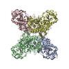
- PDB-9o6a: CryoEM structure of EcKatG S-Trp105 at 2.22 Angstrom resolution r... -
+ Open data

- Basic information
| Entry | Database: PDB / ID: 9o6a | |||||||||||||||||||||||||||
|---|---|---|---|---|---|---|---|---|---|---|---|---|---|---|---|---|---|---|---|---|---|---|---|---|---|---|---|---|
| Title | CryoEM structure of EcKatG S-Trp105 at 2.22 Angstrom resolution revealing an asymmetric sulfur center in O=S-Trp | |||||||||||||||||||||||||||
 Components Components | Catalase-peroxidase | |||||||||||||||||||||||||||
 Keywords Keywords | OXIDOREDUCTASE / Met-Tyr-Trp cofactor / heme-dependent enzyme / non-canonical amino acid | |||||||||||||||||||||||||||
| Function / homology |  Function and homology information Function and homology informationcatalase-peroxidase / catalase activity / hydrogen peroxide catabolic process / cellular response to hydrogen peroxide / heme binding / metal ion binding / cytosol Similarity search - Function | |||||||||||||||||||||||||||
| Biological species |  | |||||||||||||||||||||||||||
| Method | ELECTRON MICROSCOPY / single particle reconstruction / cryo EM / Resolution: 2.22 Å | |||||||||||||||||||||||||||
 Authors Authors | Duan, R. / Li, J. / Nathan, B. / Yang, X. / Liu, A. | |||||||||||||||||||||||||||
| Funding support |  United States, 1items United States, 1items
| |||||||||||||||||||||||||||
 Citation Citation | Journal: To Be Published Title: 2.22 Angstrom resolution cryoEM structure of EcKatG S-Trp105 with asymmetric S-oxygenation Authors: Duan, R. / Li, J. / Liu, A. | |||||||||||||||||||||||||||
| History |
|
- Structure visualization
| Structure viewer | Molecule: MolmilJmol/JSmol |
|---|
- Downloads & links
-Download
| PDBx/mmCIF format | 9o6a.cif.gz | 499.7 KB | Display | PDBx/mmCIF format |
|---|---|---|---|---|
| PDB format | pdb9o6a.ent.gz | Display | PDB format | |
| PDBx/mmJSON format | 9o6a.json.gz | Tree view | PDBx/mmJSON format | |
| Others |  Other downloads Other downloads |
-Validation report
| Arichive directory | https://data.pdbj.org/pub/pdb/validation_reports/o6/9o6aftp://data.pdbj.org/pub/pdb/validation_reports/o6/9o6a | HTTPS FTP |
|---|
-Related structure data
| Related structure data |  70168MC M: map data used to model this data C: citing same article ( |
|---|---|
| Similar structure data |
-Links
 PDBj
PDBj
- Assembly
| Deposited unit | 
|
|---|---|
| 1 |
|
-Components
| #1: Protein | Mass: 81105.672 Da / Num. of mol.: 4 Source method: isolated from a genetically manipulated source Source: (gene. exp.) #2: Chemical | ChemComp-HEM /   Mass: 616.487 Da / Num. of mol.: 4 / Source method: obtained synthetically / Formula: C34H32FeN4O4 Mass: 616.487 Da / Num. of mol.: 4 / Source method: obtained synthetically / Formula: C34H32FeN4O4#3: Water | ChemComp-HOH / |  Mass: 18.015 Da / Num. of mol.: 20 / Source method: isolated from a natural source / Formula: H2O Mass: 18.015 Da / Num. of mol.: 20 / Source method: isolated from a natural source / Formula: H2OHas ligand of interest | Y | Has protein modification | Y | |
|---|
-Experimental details
-Experiment
| Experiment | Method: ELECTRON MICROSCOPY |
|---|---|
| EM experiment | Aggregation state: PARTICLE / 3D reconstruction method: single particle reconstruction |
- Sample preparation
| Component | Name: EcKatG S-Trp105 / Type: COMPLEX Details: KatG protein with 3-benzothienyl-L-alanine (S-Trp) genetically incorporated in place of W105 Entity ID: #1 / Source: RECOMBINANT | |||||||||||||||
|---|---|---|---|---|---|---|---|---|---|---|---|---|---|---|---|---|
| Molecular weight | Value: 0.32 MDa / Experimental value: NO | |||||||||||||||
| Source (natural) | Organism: | |||||||||||||||
| Source (recombinant) | Organism: | |||||||||||||||
| Buffer solution | pH: 8 / Details: 50 mM Tris-HCl, 50 mM NaCl at pH 8 | |||||||||||||||
| Buffer component |
| |||||||||||||||
| Specimen | Conc.: 0.12 mg/ml / Embedding applied: NO / Shadowing applied: NO / Staining applied: NO / Vitrification applied: YES Details: The KatG S-Trp105 was dissolved in the 50 mM Tris-HCl, 50 mM NaCl buffer at pH 8 | |||||||||||||||
| Specimen support | Grid material: COPPER / Grid mesh size: 200 divisions/in. / Grid type: Quantifoil R2/1 | |||||||||||||||
| Vitrification | Instrument: FEI VITROBOT MARK IV / Cryogen name: ETHANE / Humidity: 100 % / Chamber temperature: 277 K Details: 3 microliter of the sample was applied to each grid and blotted for 3 s |
- Electron microscopy imaging
| Experimental equipment |  Model: Titan Krios / Image courtesy: FEI Company |
|---|---|
| Microscopy | Model: TFS KRIOS |
| Electron gun | Electron source:  FIELD EMISSION GUN / Accelerating voltage: 300 kV / Illumination mode: FLOOD BEAM FIELD EMISSION GUN / Accelerating voltage: 300 kV / Illumination mode: FLOOD BEAM |
| Electron lens | Mode: BRIGHT FIELD / Nominal magnification: 130000 X / Nominal defocus max: 2000 nm / Nominal defocus min: 1000 nm |
| Specimen holder | Cryogen: NITROGEN / Specimen holder model: FEI TITAN KRIOS AUTOGRID HOLDER |
| Image recording | Average exposure time: 6.58 sec. / Electron dose: 50 e/Å2 / Film or detector model: TFS FALCON 4i (4k x 4k) |
| EM imaging optics | Energyfilter name: TFS Selectris X / Energyfilter slit width: 10 eV |
- Processing
| EM software |
| |||||||||||||||||||||||||||||||||||||||||||||
|---|---|---|---|---|---|---|---|---|---|---|---|---|---|---|---|---|---|---|---|---|---|---|---|---|---|---|---|---|---|---|---|---|---|---|---|---|---|---|---|---|---|---|---|---|---|---|
| CTF correction | Type: PHASE FLIPPING AND AMPLITUDE CORRECTION | |||||||||||||||||||||||||||||||||||||||||||||
| Particle selection | Num. of particles selected: 7762366 / Details: Particles were selected from blob picking. | |||||||||||||||||||||||||||||||||||||||||||||
| Symmetry | Point symmetry: D4 (2x4 fold dihedral) | |||||||||||||||||||||||||||||||||||||||||||||
| 3D reconstruction | Resolution: 2.22 Å / Resolution method: FSC 0.143 CUT-OFF / Num. of particles: 884126 / Algorithm: FOURIER SPACE / Num. of class averages: 43 / Symmetry type: POINT | |||||||||||||||||||||||||||||||||||||||||||||
| Atomic model building | B value: 7.92 / Protocol: AB INITIO MODEL / Space: REAL | |||||||||||||||||||||||||||||||||||||||||||||
| Atomic model building | PDB-ID: 7JZ6 Pdb chain-ID: A / Accession code: 7JZ6 / Source name: PDB / Type: experimental model | |||||||||||||||||||||||||||||||||||||||||||||
| Refinement | Stereochemistry target values: REAL-SPACE (WEIGHTED MAP SUM AT ATOM CENTERS) | |||||||||||||||||||||||||||||||||||||||||||||
| Refine LS restraints |
|