Movie
Movie Controller

[English] 日本語
 Yorodumi
- EMDB-70168: CryoEM structure of EcKatG S-Trp105 at 2.22 Angstrom resolution r... -
+ Open data

- Basic information
| Entry |  | |||||||||
|---|---|---|---|---|---|---|---|---|---|---|
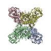
| Title | CryoEM structure of EcKatG S-Trp105 at 2.22 Angstrom resolution revealing an asymmetric sulfur center in O=S-Trp | |||||||||
 Map data Map data | Density modified map at 2.22 angstrom resolution | |||||||||
 Sample Sample |
| |||||||||
 Keywords Keywords | Met-Tyr-Trp cofactor / heme-dependent enzyme / non-canonical amino acid / OXIDOREDUCTASE | |||||||||
| Function / homology |  Function and homology information Function and homology informationcatalase-peroxidase / catalase activity / hydrogen peroxide catabolic process / cellular response to hydrogen peroxide / heme binding / metal ion binding / cytosol Similarity search - Function | |||||||||
| Biological species |  | |||||||||
| Method | single particle reconstruction / cryo EM / Resolution: 2.22 Å | |||||||||
 Authors Authors | Duan R / Li J / Nathan B / Yang X / Liu A | |||||||||
| Funding support |  United States, 1 items United States, 1 items
| |||||||||
 Citation Citation | Journal: To Be Published Title: 2.22 Angstrom resolution cryoEM structure of EcKatG S-Trp105 with asymmetric S-oxygenation Authors: Duan R / Li J / Liu A | |||||||||
| History |
|
- Structure visualization
| Supplemental images |
|---|

- Downloads & links
-EMDB archive
| Map data | emd_70168.map.gz | 5.4 MB | EMDB map data format | |
|---|---|---|---|---|
| Header (meta data) | emd-70168-v30.xmlemd-70168.xml | 23.4 KB 23.4 KB | Display Display | EMDB header |
| FSC (resolution estimation) | emd_70168_fsc.xml | 10.9 KB | Display | FSC data file |
| Images |  emd_70168.png emd_70168.png | 99.7 KB | ||
| Filedesc metadata | emd-70168.cif.gz | 7.2 KB | ||
| Others | emd_70168_additional_1.map.gzemd_70168_half_map_1.map.gzemd_70168_half_map_2.map.gz | 26.4 MB 48.9 MB 48.9 MB | ||
| Archive directory |  http://ftp.pdbj.org/pub/emdb/structures/EMD-70168ftp://ftp.pdbj.org/pub/emdb/structures/EMD-70168 http://ftp.pdbj.org/pub/emdb/structures/EMD-70168ftp://ftp.pdbj.org/pub/emdb/structures/EMD-70168 | HTTPS FTP |
-Related structure data
| Related structure data |  9o6aMC M: atomic model generated by this map C: citing same article ( |
|---|---|
| Similar structure data |
-Links
| EMDB pages | EMDB (EBI/PDBe) / EMDataResource |
|---|---|
| Related items in Molecule of the Month |

-Map
| File | Download / File: emd_70168.map.gz / Format: CCP4 / Size: 52.7 MB / Type: IMAGE STORED AS FLOATING POINT NUMBER (4 BYTES) | ||||||||||||||||||||||||||||||||||||
|---|---|---|---|---|---|---|---|---|---|---|---|---|---|---|---|---|---|---|---|---|---|---|---|---|---|---|---|---|---|---|---|---|---|---|---|---|---|
| Annotation | Density modified map at 2.22 angstrom resolution | ||||||||||||||||||||||||||||||||||||
| Projections & slices | Image control
Images are generated by Spider. | ||||||||||||||||||||||||||||||||||||
| Voxel size | X=Y=Z: 0.954 Å | ||||||||||||||||||||||||||||||||||||
| Density |
| ||||||||||||||||||||||||||||||||||||
| Symmetry | Space group: 1 | ||||||||||||||||||||||||||||||||||||
| Details | EMDB XML:
|
 X (Sec.)
 Y (Row.)
 Z (Col.)


-Supplemental data
-Additional map: Unsharpened map from cryoSPARC
| File | emd_70168_additional_1.map | ||||||||||||
|---|---|---|---|---|---|---|---|---|---|---|---|---|---|
| Annotation | Unsharpened map from cryoSPARC | ||||||||||||
| Projections & Slices |
| ||||||||||||
| Density Histograms |


-Half map: Unsharpened half-map from cryoSPARC
| File | emd_70168_half_map_1.map | ||||||||||||
|---|---|---|---|---|---|---|---|---|---|---|---|---|---|
| Annotation | Unsharpened half-map from cryoSPARC | ||||||||||||
| Projections & Slices |
| ||||||||||||
| Density Histograms |


-Half map: Unsharpened half-map from cryoSPARC
| File | emd_70168_half_map_2.map | ||||||||||||
|---|---|---|---|---|---|---|---|---|---|---|---|---|---|
| Annotation | Unsharpened half-map from cryoSPARC | ||||||||||||
| Projections & Slices |
| ||||||||||||
| Density Histograms |


- Sample components
-Entire : EcKatG S-Trp105
| Entire | Name: EcKatG S-Trp105 |
|---|---|
| Components |
|
-Supramolecule #1: EcKatG S-Trp105
| Supramolecule | Name: EcKatG S-Trp105 / type: complex / ID: 1 / Parent: 0 / Macromolecule list: #1 Details: KatG protein with 3-benzothienyl-L-alanine (S-Trp) genetically incorporated in place of W105 |
|---|---|
| Source (natural) | Organism: |
| Molecular weight | Theoretical: 320 KDa |
-Macromolecule #1: Catalase-peroxidase
| Macromolecule | Name: Catalase-peroxidase / type: protein_or_peptide / ID: 1 / Number of copies: 4 / Enantiomer: LEVO / EC number: catalase-peroxidase |
|---|---|
| Source (natural) | Organism: |
| Molecular weight | Theoretical: 81.105672 KDa |
| Recombinant expression | Organism: |
| Sequence | String: MSTSDDIHNT TATGKCPFHQ GGHDQSAGAG TTTRDWWPNQ LRVDLLNQHS NRSNPLGEDF DYRKEFSKLD YYGLKKDLKA LLTESQPWW PADWGSYAGL FIRMA(A1B9V)HGAG TYRSIDGRGG AGRGQQRFAP LNSWPDNVSL DKARRLLWPI KQKYG QKIS ...String: MSTSDDIHNT TATGKCPFHQ GGHDQSAGAG TTTRDWWPNQ LRVDLLNQHS NRSNPLGEDF DYRKEFSKLD YYGLKKDLKA LLTESQPWW PADWGSYAGL FIRMA(A1B9V)HGAG TYRSIDGRGG AGRGQQRFAP LNSWPDNVSL DKARRLLWPI KQKYG QKIS WADLFILAGN VALENSGFRT FGFGAGREDV WEPDLDVNWG DEKAWLTHRH PEALAKAPLG ATEMGLIYVN PEGPDH SGE PLSAAAAIRA TFGNMGMNDE ETVALIAGGH TLGKTHGAGP TSNVGPDPEA APIEEQGLGW ASTYGSGVGA DAITSGL EV VWTQTPTQWS NYFFENLFKY EWVQTRSPAG AIQFEAVDAP EIIPDPFDPS KKRKPTMLVT DLTLRFDPEF EKISRRFL N DPQAFNEAFA RAWFKLTHRD MGPKSRYIGP EVPKEDLIWQ DPLPQPIYNP TEQDIIDLKF AIADSGLSVS ELVSVAWAS ASTFRGGDKR GGANGARLAL MPQRDWDVNA AAVRALPVLE KIQKESGKAS LADIIVLAGV VGVEKAASAA GLSIHVPFAP GRVDARQDQ TDIEMFELLE PIADGFRNYR ARLDVSTTES LLIDKAQQLT LTAPEMTALV GGMRVLGANF DGSKNGVFTD R VGVLSNDF FVNLLDMRYE WKATDESKEL FEGRDRETGE VKFTASRADL VFGSNSVLRA VAEVYASSDA HEKFVKDFVA AW VKVMNLD RFDLLEHHHH HH UniProtKB: Catalase-peroxidase |
-Macromolecule #2: PROTOPORPHYRIN IX CONTAINING FE
| Macromolecule | Name: PROTOPORPHYRIN IX CONTAINING FE / type: ligand / ID: 2 / Number of copies: 4 / Formula: HEM |
|---|---|
| Molecular weight | Theoretical: 616.487 Da |
| Chemical component information |  ChemComp-HEM: |
-Macromolecule #3: water
| Macromolecule | Name: water / type: ligand / ID: 3 / Number of copies: 20 / Formula: HOH |
|---|---|
| Molecular weight | Theoretical: 18.015 Da |
| Chemical component information |  ChemComp-HOH: |
-Experimental details
-Structure determination
| Method | cryo EM |
|---|---|
 Processing Processing | single particle reconstruction |
| Aggregation state | particle |
-Sample preparation
| Concentration | 0.12 mg/mL | |||||||||
|---|---|---|---|---|---|---|---|---|---|---|
| Buffer | pH: 8 Component:
Details: 50 mM Tris-HCl, 50 mM NaCl at pH 8 | |||||||||
| Grid | Model: Quantifoil R2/1 / Material: COPPER / Mesh: 200 / Pretreatment - Type: GLOW DISCHARGE / Pretreatment - Time: 30 sec. / Pretreatment - Atmosphere: OTHER / Pretreatment - Pressure: 0.026000000000000002 kPa | |||||||||
| Vitrification | Cryogen name: ETHANE / Chamber humidity: 100 % / Chamber temperature: 277 K / Instrument: FEI VITROBOT MARK IV Details: 3 microliter of the sample was applied to each grid and blotted for 3 s. | |||||||||
| Details | The KatG S-Trp105 was dissolved in the 50 mM Tris-HCl, 50 mM NaCl buffer at pH 8 |
- Electron microscopy
| Microscope | TFS KRIOS |
|---|---|
| Specialist optics | Energy filter - Name: TFS Selectris X / Energy filter - Slit width: 10 eV |
| Image recording | Film or detector model: TFS FALCON 4i (4k x 4k) / Average exposure time: 6.58 sec. / Average electron dose: 50.0 e/Å2 |
| Electron beam | Acceleration voltage: 300 kV / Electron source:  FIELD EMISSION GUN FIELD EMISSION GUN |
| Electron optics | Illumination mode: FLOOD BEAM / Imaging mode: BRIGHT FIELD / Nominal defocus max: 2.0 µm / Nominal defocus min: 1.0 µm / Nominal magnification: 130000 |
| Sample stage | Specimen holder model: FEI TITAN KRIOS AUTOGRID HOLDER / Cooling holder cryogen: NITROGEN |
| Experimental equipment |  Model: Titan Krios / Image courtesy: FEI Company |
+Image processing

-Atomic model buiding 1
| Initial model | PDB ID: Chain - Chain ID: A / Chain - Source name: PDB / Chain - Initial model type: experimental model |
|---|---|
| Refinement | Space: REAL / Protocol: AB INITIO MODEL / Overall B value: 7.92 |
| Output model | PDB-9o6a: |