Movie
Movie Controller


+ Open data

- Basic information
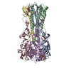
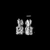
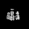
| Entry | Database: PDB / ID: 9n8p | |||||||||
|---|---|---|---|---|---|---|---|---|---|---|
| Title | Subtomogram average of dimers of influenza HA trimers | |||||||||
 Components Components |
| |||||||||
 Keywords Keywords | VIRAL PROTEIN / Hemagglutinin | |||||||||
| Function / homology |  Function and homology information Function and homology informationTransport of HA trimer, NA tetramer and M2 tetramer from the endoplasmic reticulum to the Golgi Apparatus / Assembly of Viral Components at the Budding Site / Influenza Infection / Fusion of the Influenza Virion to the Host Cell Endosome / Release / Budding / Packaging of Eight RNA Segments / Uncoating of the Influenza Virion / Entry of Influenza Virion into Host Cell via Endocytosis / Viral mRNA Translation ...Transport of HA trimer, NA tetramer and M2 tetramer from the endoplasmic reticulum to the Golgi Apparatus / Assembly of Viral Components at the Budding Site / Influenza Infection / Fusion of the Influenza Virion to the Host Cell Endosome / Release / Budding / Packaging of Eight RNA Segments / Uncoating of the Influenza Virion / Entry of Influenza Virion into Host Cell via Endocytosis / Viral mRNA Translation / viral budding from plasma membrane / Immunoregulatory interactions between a Lymphoid and a non-Lymphoid cell / clathrin-dependent endocytosis of virus by host cell / host cell surface receptor binding / fusion of virus membrane with host plasma membrane / fusion of virus membrane with host endosome membrane / viral envelope / virion attachment to host cell / host cell plasma membrane / virion membrane / extracellular region / plasma membrane Similarity search - Function | |||||||||
| Biological species |   Influenza A virus Influenza A virus | |||||||||
| Method | ELECTRON MICROSCOPY / subtomogram averaging / cryo EM / Resolution: 9 Å | |||||||||
 Authors Authors | Huang, Q.J. / Song, K. / Schiffer, C.A. / Somasundaran, M. | |||||||||
| Funding support |  United States, 2items United States, 2items
| |||||||||
 Citation Citation | Journal: Proc Natl Acad Sci U S A / Year: 2025 Title: Virion-associated influenza hemagglutinin clusters upon sialic acid binding visualized by cryoelectron tomography. Authors: Qiuyu J Huang / Ryan Kim / Kangkang Song / Nikolaus Grigorieff / James B Munro / Celia A Schiffer / Mohan Somasundaran / Abstract: Influenza viruses are enveloped, negative-sense single-stranded RNA viruses covered in a dense layer of glycoproteins. Hemagglutinin (HA) accounts for 80 to 90% of influenza glycoprotein and plays a ...Influenza viruses are enveloped, negative-sense single-stranded RNA viruses covered in a dense layer of glycoproteins. Hemagglutinin (HA) accounts for 80 to 90% of influenza glycoprotein and plays a role in host cell binding and membrane fusion. While previous studies have characterized structures of purified receptor-free and receptor-bound HA, the effect of receptor binding on HA organization and structure on virions remains unknown. Here, we used cryoelectron tomography to visualize influenza virions bound to a sialic acid receptor mimic. Overall, receptor binding did not result in significant changes in viral morphology; however, we observed rearrangements of HA trimer organization and orientation. Compared to the even interglycoprotein spacing of unliganded HA trimers, receptor binding promotes HA trimer clustering and the formation of a triplet of trimers. Subtomogram averaging and refinement yielded 8 to 10 Å reconstructions that allowed us to visualize specific contacts between HAs from neighboring trimers and identify molecular features that mediate clustering. Taken together, we present structural evidence that receptor binding triggers clustering of HA trimers, revealing an additional layer of HA dynamics and plasticity. | |||||||||
| History |
|
- Structure visualization
| Structure viewer | Molecule: MolmilJmol/JSmol |
|---|
- Downloads & links
-Download
| PDBx/mmCIF format | 9n8p.cif.gz | 506.8 KB | Display | PDBx/mmCIF format |
|---|---|---|---|---|
| PDB format | pdb9n8p.ent.gz | 426.3 KB | Display | PDB format |
| PDBx/mmJSON format | 9n8p.json.gz | Tree view | PDBx/mmJSON format | |
| Others |  Other downloads Other downloads |
-Validation report
| Arichive directory | https://data.pdbj.org/pub/pdb/validation_reports/n8/9n8pftp://data.pdbj.org/pub/pdb/validation_reports/n8/9n8p | HTTPS FTP |
|---|
-Related structure data
| Related structure data |  47030MC M: map data used to model this data C: citing same article ( |
|---|---|
| Similar structure data |
-Links
 PDBj
PDBj


- Assembly
| Deposited unit | 
|
|---|---|
| 1 |
|
-Components
| #1: Protein | Mass: 36733.402 Da / Num. of mol.: 6 / Source method: isolated from a natural source Source: (natural) Influenza A virus (A/Puerto Rico/8/1934(H1N1))Strain: A/Puerto Rico/8/1934 / References: UniProt: P03452 #2: Protein | Mass: 18242.221 Da / Num. of mol.: 6 / Source method: isolated from a natural source Source: (natural) Influenza A virus (A/Puerto Rico/8/1934(H1N1))Strain: A/Puerto Rico/8/1934 / References: UniProt: P03452 #3: Polysaccharide | N-acetyl-alpha-neuraminic acid-(2-6)-beta-D-galactopyranose Source method: isolated from a genetically manipulated source Has ligand of interest | Y | Has protein modification | Y | |
|---|
-Experimental details
-Experiment
| Experiment | Method: ELECTRON MICROSCOPY |
|---|---|
| EM experiment | Aggregation state: PARTICLE / 3D reconstruction method: subtomogram averaging |
- Sample preparation
| Component | Name: Influenza A virus (A/Puerto Rico/8/1934(H1N1)) / Type: VIRUS Details: Virus produced in embryonated eggs and purified from allantoic fluid. Entity ID: #1-#2 / Source: NATURAL |
|---|---|
| Source (natural) | Organism: Influenza A virus (A/Puerto Rico/8/1934(H1N1)) / Strain: A/Puerto Rico/8/1934 |
| Details of virus | Empty: NO / Enveloped: YES / Isolate: STRAIN / Type: VIRION |
| Buffer solution | pH: 7.4 |
| Specimen | Conc.: 1 mg/ml / Embedding applied: NO / Shadowing applied: NO / Staining applied: NO / Vitrification applied: YES Details: Virus was incubated with Sialylneolacto-N-tetraose c (LSTc) at a final concentration of 100 um |
| Specimen support | Grid material: COPPER / Grid mesh size: 200 divisions/in. / Grid type: Quantifoil R2/2 |
| Vitrification | Instrument: EMS-002 RAPID IMMERSION FREEZER / Cryogen name: ETHANE-PROPANE |
- Electron microscopy imaging
| Experimental equipment |  Model: Titan Krios / Image courtesy: FEI Company |
|---|---|
| Microscopy | Model: TFS KRIOS |
| Electron gun | Electron source:  FIELD EMISSION GUN / Accelerating voltage: 300 kV / Illumination mode: FLOOD BEAM FIELD EMISSION GUN / Accelerating voltage: 300 kV / Illumination mode: FLOOD BEAM |
| Electron lens | Mode: BRIGHT FIELD / Nominal magnification: 42000 X / Nominal defocus max: 6000 nm / Nominal defocus min: 3000 nm / Cs: 2.7 mm |
| Specimen holder | Cryogen: NITROGEN |
| Image recording | Electron dose: 1.86 e/Å2 / Avg electron dose per subtomogram: 120 e/Å2 / Film or detector model: GATAN K3 (6k x 4k) |
- Processing
| EM software |
| ||||||||||||||||||||||||
|---|---|---|---|---|---|---|---|---|---|---|---|---|---|---|---|---|---|---|---|---|---|---|---|---|---|
| CTF correction | Type: PHASE FLIPPING AND AMPLITUDE CORRECTION | ||||||||||||||||||||||||
| Symmetry | Point symmetry: C1 (asymmetric) | ||||||||||||||||||||||||
| 3D reconstruction | Resolution: 9 Å / Resolution method: FSC 0.143 CUT-OFF / Num. of particles: 9283 / Symmetry type: POINT | ||||||||||||||||||||||||
| EM volume selection | Num. of tomograms: 16 / Num. of volumes extracted: 26500 | ||||||||||||||||||||||||
| Atomic model building | Protocol: FLEXIBLE FIT / Space: REAL Details: Rigid body docking was performed in ChimeraX. Phenix was used for flexible real space refinement and Schrodinger Protein Preparation Wizard was used to perform final energy minimization. | ||||||||||||||||||||||||
| Atomic model building | PDB-ID: 1RVZ Accession code: 1RVZ / Source name: PDB / Type: experimental model |