 Movie
Movie Controller

[English] 日本語
 Yorodumi
- PDB-9n37: Cryo-EM structure of Rubisco from Arabidopsis thaliana with the 1... -
+ Open data

- Basic information
| Entry | Database: PDB / ID: 9n37 | ||||||||||||||||||||||||
|---|---|---|---|---|---|---|---|---|---|---|---|---|---|---|---|---|---|---|---|---|---|---|---|---|---|
| Title | Cryo-EM structure of Rubisco from Arabidopsis thaliana with the 1A small subunit isoform | ||||||||||||||||||||||||
 Components Components |
| ||||||||||||||||||||||||
 Keywords Keywords | PHOTOSYNTHESIS / carboxylase / oxygenase / TIM barrel | ||||||||||||||||||||||||
| Function / homology |  Function and homology information Function and homology informationsalicylic acid binding / thylakoid lumen / ribulose bisphosphate carboxylase complex assembly / chloroplast membrane / plant-type cell wall / photorespiration / ribulose-bisphosphate carboxylase / ribulose-bisphosphate carboxylase activity / apoplast / thylakoid ...salicylic acid binding / thylakoid lumen / ribulose bisphosphate carboxylase complex assembly / chloroplast membrane / plant-type cell wall / photorespiration / ribulose-bisphosphate carboxylase / ribulose-bisphosphate carboxylase activity / apoplast / thylakoid / reductive pentose-phosphate cycle / response to abscisic acid / chloroplast envelope / chloroplast stroma / plastid / chloroplast thylakoid membrane / response to cadmium ion / response to cold / photosynthesis / monooxygenase activity / chloroplast / cytosolic ribosome / copper ion binding / mRNA binding / magnesium ion binding / mitochondrion Similarity search - Function | ||||||||||||||||||||||||
| Biological species |   Arabidopsis thaliana (thale cress) Arabidopsis thaliana (thale cress) | ||||||||||||||||||||||||
| Method | ELECTRON MICROSCOPY / single particle reconstruction / cryo EM / Resolution: 2.2 Å | ||||||||||||||||||||||||
 Authors Authors | Stavros, A. / Askey, B. / Ceminsky, M. / Gunn, L.H. | ||||||||||||||||||||||||
| Funding support |  Sweden, Sweden,  United States, 3items United States, 3items
| ||||||||||||||||||||||||
 Citation Citation | Journal: Proc Natl Acad Sci U S A / Year: 2026 Title: Rubisco kinetic acclimation at the holoenzyme level. Authors: Bryce Askey / Maddie Ceminsky / Elena Scott / Yongsheng Wang / Zhen Guo Oh / Stavros Azinas / Arthur Laganowsky / Laura Helen Gunn / Abstract: In plants, the CO-fixing enzyme Rubisco is hexadecameric, with each mature holoenzyme containing eight small subunits (SSus). Many plants express multiple SSus and vary their expression in response ...In plants, the CO-fixing enzyme Rubisco is hexadecameric, with each mature holoenzyme containing eight small subunits (SSus). Many plants express multiple SSus and vary their expression in response to environmental cues. Previous work indicates that this may allow fine-tuning of Rubisco's performance in a variable environment (i.e., kinetic acclimation). Despite SSu pools being heterogeneous and dynamic, nearly no evidence exists for holoenzyme-level heterogeneity. Here, we characterized the structural and kinetic plasticity of Rubisco. We first established that SSu-heterogeneous Rubisco exists in and quantified the prevalence of heterogeneity. We found SSu-heterogeneous Rubisco to make up over half of the Rubisco pool when heterologously expressed. This Rubisco contained at least four unique SSu ratios, indicating a variety of holoenzyme arrangements are possible. We then tested the kinetic effect of different SSus and found heterogeneity to have an antagonistic effect on substrate and inhibitor affinity. Kinetic differences between the SSus correlated with changes in local flexibility, and cryo-EM analysis illustrated a structural mechanism through which SSus may influence catalysis. Our kinetic and structural findings align with the hypothesized role of SSus in kinetic acclimation, as we observed the warm temperature-expressed SSu of to confer a stabilizing effect to the active site relative to the cool temperature-expressed SSu. This increase in stability manifested as a reduction in flexibility and increase in substrate affinity, indicating that fine-tuning of local stability may underlie Rubisco kinetic acclimation. | ||||||||||||||||||||||||
| History |
|
- Structure visualization
| Structure viewer | Molecule: MolmilJmol/JSmol |
|---|
- Downloads & links
-Download
| PDBx/mmCIF format | 9n37.cif.gz | 1.9 MB | Display | PDBx/mmCIF format |
|---|---|---|---|---|
| PDB format | pdb9n37.ent.gz | 1.3 MB | Display | PDB format |
| PDBx/mmJSON format | 9n37.json.gz | Tree view | PDBx/mmJSON format | |
| Others |  Other downloads Other downloads |
-Validation report
| Arichive directory | https://data.pdbj.org/pub/pdb/validation_reports/n3/9n37ftp://data.pdbj.org/pub/pdb/validation_reports/n3/9n37 | HTTPS FTP |
|---|
-Related structure data
| Related structure data |  48847MC  9murC C: citing same article ( M: map data used to model this data |
|---|---|
| Similar structure data |

-Links
 PDBj
PDBj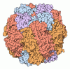
- Assembly
| Deposited unit | 
|
|---|---|
| 1 |
|
-Components
| #1: Protein | Mass: 53063.883 Da / Num. of mol.: 8 Source method: isolated from a genetically manipulated source Source: (gene. exp.) Arabidopsis thaliana (thale cress) / Gene: rbcL, AtCg00490Production host:   Escherichia coli 'BL21-Gold(DE3)pLysS AG' (bacteria) Escherichia coli 'BL21-Gold(DE3)pLysS AG' (bacteria)References: UniProt: O03042, ribulose-bisphosphate carboxylase #2: Protein | Mass: 15544.694 Da / Num. of mol.: 8 Source method: isolated from a genetically manipulated source Source: (gene. exp.) Arabidopsis thaliana (thale cress) / Gene: RBCS-1A, ATS1A, At1g67090, F1O19.14, F5A8.1Production host: Escherichia coli 'BL21-Gold(DE3)pLysS AG' (bacteria)References: UniProt: P10795 #3: Chemical | ChemComp-MG /   Mass: 24.305 Da / Num. of mol.: 8 / Source method: obtained synthetically / Formula: Mg / Feature type: SUBJECT OF INVESTIGATION Mass: 24.305 Da / Num. of mol.: 8 / Source method: obtained synthetically / Formula: Mg / Feature type: SUBJECT OF INVESTIGATIONHas ligand of interest | Y | Has protein modification | Y | |
|---|
-Experimental details
-Experiment
| Experiment | Method: ELECTRON MICROSCOPY |
|---|---|
| EM experiment | Aggregation state: PARTICLE / 3D reconstruction method: single particle reconstruction |
- Sample preparation
| Component |
| ||||||||||||||||||||||||
|---|---|---|---|---|---|---|---|---|---|---|---|---|---|---|---|---|---|---|---|---|---|---|---|---|---|
| Molecular weight |
| ||||||||||||||||||||||||
| Source (natural) |
| ||||||||||||||||||||||||
| Source (recombinant) |
| ||||||||||||||||||||||||
| Buffer solution | pH: 8 | ||||||||||||||||||||||||
| Specimen | Conc.: 1 mg/ml / Embedding applied: NO / Shadowing applied: NO / Staining applied: NO / Vitrification applied: YES | ||||||||||||||||||||||||
| Vitrification | Instrument: FEI VITROBOT MARK IV / Cryogen name: ETHANE / Humidity: 100 % / Chamber temperature: 277.15 K |
- Electron microscopy imaging
| Experimental equipment |  Model: Titan Krios / Image courtesy: FEI Company |
|---|---|
| Microscopy | Model: TFS KRIOS |
| Electron gun | Electron source:  FIELD EMISSION GUN / Accelerating voltage: 300 kV / Illumination mode: SPOT SCAN FIELD EMISSION GUN / Accelerating voltage: 300 kV / Illumination mode: SPOT SCAN |
| Electron lens | Mode: BRIGHT FIELD / Nominal magnification: 130000 X / Nominal defocus max: 2400 nm / Nominal defocus min: 600 nm |
| Specimen holder | Cryogen: NITROGEN |
| Image recording | Electron dose: 40 e/Å2 / Film or detector model: GATAN K3 (6k x 4k) |
- Processing
| EM software | Name: PHENIX / Version: 1.21.1_5286 / Category: model refinement | ||||||||||||||||||||||||
|---|---|---|---|---|---|---|---|---|---|---|---|---|---|---|---|---|---|---|---|---|---|---|---|---|---|
| CTF correction | Type: PHASE FLIPPING AND AMPLITUDE CORRECTION | ||||||||||||||||||||||||
| Particle selection | Num. of particles selected: 491616 | ||||||||||||||||||||||||
| Symmetry | Point symmetry: D4 (2x4 fold dihedral) | ||||||||||||||||||||||||
| 3D reconstruction | Resolution: 2.2 Å / Resolution method: FSC 0.143 CUT-OFF / Num. of particles: 256188 / Symmetry type: POINT | ||||||||||||||||||||||||
| Atomic model building | Protocol: RIGID BODY FIT / Space: REAL | ||||||||||||||||||||||||
| Atomic model building | PDB-ID: 5IU0 Accession code: 5IU0 / Source name: PDB / Type: experimental model | ||||||||||||||||||||||||
| Refinement | Cross valid method: NONE Stereochemistry target values: GeoStd + Monomer Library + CDL v1.2 | ||||||||||||||||||||||||
| Displacement parameters | Biso mean: 83.71 Å2 | ||||||||||||||||||||||||
| Refine LS restraints |
|

