 Movie
Movie Controller


+ Open data

- Basic information
| Entry | Database: PDB / ID: 9n0x | ||||||||||||||||||||||||
|---|---|---|---|---|---|---|---|---|---|---|---|---|---|---|---|---|---|---|---|---|---|---|---|---|---|
| Title | Cryo-EM structure of human PSS2 | ||||||||||||||||||||||||
 Components Components | Phosphatidylserine synthase 2 | ||||||||||||||||||||||||
 Keywords Keywords | MEMBRANE PROTEIN / PS lipid | ||||||||||||||||||||||||
| Function / homology |  Function and homology information Function and homology informationCDP-diacylglycerol-serine O-phosphatidyltransferase activity / L-serine-phosphatidylethanolamine phosphatidyltransferase / L-serine-phosphatidylethanolamine phosphatidyltransferase activity / Synthesis of PS / phosphatidylserine biosynthetic process / transferase activity / endoplasmic reticulum membrane / membrane Similarity search - Function | ||||||||||||||||||||||||
| Biological species |  Homo sapiens (human) Homo sapiens (human) | ||||||||||||||||||||||||
| Method | ELECTRON MICROSCOPY / single particle reconstruction / cryo EM / Resolution: 3.3 Å | ||||||||||||||||||||||||
 Authors Authors | Li, D.Y. / Li, X.C. | ||||||||||||||||||||||||
| Funding support |  United States, 1items United States, 1items
| ||||||||||||||||||||||||
 Citation Citation | Journal: Proc Natl Acad Sci U S A / Year: 2025 Title: Molecular insights into human phosphatidylserine synthase 2 and its regulation of SREBP pathways. Authors: Dongyu Li / Hongwen Chen / Goncalo Vale / Nadia Elghobashi-Meinhardt / Alexandra Hatton / Shunxing Rong / Jeffrey G McDonald / Xiaochun Li /  Abstract: Homologous proteins share similar sequences, enabling them to work together in cells to support normal physiological functions. Phosphatidylserine synthases 1 and 2 (PSS1 and PSS2) are homologous ...Homologous proteins share similar sequences, enabling them to work together in cells to support normal physiological functions. Phosphatidylserine synthases 1 and 2 (PSS1 and PSS2) are homologous enzymes that catalyze the synthesis of phosphatidylserine (PS) from different substrates. PSS2 shows a preference for phosphatidylethanolamine (PE) as its substrate, whereas PSS1 can utilize either PE or phosphatidylcholine. Previous studies showed that inhibiting PSS1 promotes SREBP-2 cleavage. Interestingly, despite their homology, our findings reveal that PSS2 exerts an opposing effect on the cleavage of both SREBP-1 and SREBP-2. We resolved the cryo-electron microscopy (cryo-EM) structure of human PSS2 at 3.3 Å resolution. Structural comparison of the catalytic cavities between PSS1 and PSS2 along with molecular dynamics simulations uncovers the molecular details behind the substrate preference of PSS2 for PE. The lipidomic analysis showed that PSS2 deficiency leads to PE accumulation in the endoplasmic reticulum, which has been shown to inhibit the cleavage of sterol regulatory element-binding proteins (SREBPs) in mice. Thus, our findings reveal the intricate network of intracellular phospholipid metabolism and underscore the distinct regulatory roles of homologous proteins in cellular activities. | ||||||||||||||||||||||||
| History |
|
- Structure visualization
| Structure viewer | Molecule: MolmilJmol/JSmol |
|---|
- Downloads & links
-Download
| PDBx/mmCIF format | 9n0x.cif.gz | 175.9 KB | Display | PDBx/mmCIF format |
|---|---|---|---|---|
| PDB format | pdb9n0x.ent.gz | 136 KB | Display | PDB format |
| PDBx/mmJSON format | 9n0x.json.gz | Tree view | PDBx/mmJSON format | |
| Others |  Other downloads Other downloads |
-Validation report
| Arichive directory | https://data.pdbj.org/pub/pdb/validation_reports/n0/9n0xftp://data.pdbj.org/pub/pdb/validation_reports/n0/9n0x | HTTPS FTP |
|---|
-Related structure data
| Related structure data |  48797MC M: map data used to model this data C: citing same article ( |
|---|---|
| Similar structure data |
-Links
 PDBj
PDBj- Assembly
| Deposited unit | 
|
|---|---|
| 1 |
|
-Components
| #1: Protein | Mass: 56308.660 Da / Num. of mol.: 2 Source method: isolated from a genetically manipulated source Source: (gene. exp.) Homo sapiens (human) / Gene: PTDSS2, PSS2 / Production host: Homo sapiens (human)References: UniProt: Q9BVG9, L-serine-phosphatidylethanolamine phosphatidyltransferase #2: Chemical | ChemComp-P5S /   Mass: 792.075 Da / Num. of mol.: 4 / Source method: obtained synthetically / Formula: C42H82NO10P / Feature type: SUBJECT OF INVESTIGATION Mass: 792.075 Da / Num. of mol.: 4 / Source method: obtained synthetically / Formula: C42H82NO10P / Feature type: SUBJECT OF INVESTIGATION#3: Chemical |   Mass: 40.078 Da / Num. of mol.: 2 / Source method: obtained synthetically / Formula: Ca / Feature type: SUBJECT OF INVESTIGATION Mass: 40.078 Da / Num. of mol.: 2 / Source method: obtained synthetically / Formula: Ca / Feature type: SUBJECT OF INVESTIGATION#4: Chemical | 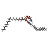  Mass: 715.981 Da / Num. of mol.: 2 / Source method: obtained synthetically / Formula: C39H74NO8P / Feature type: SUBJECT OF INVESTIGATION Mass: 715.981 Da / Num. of mol.: 2 / Source method: obtained synthetically / Formula: C39H74NO8P / Feature type: SUBJECT OF INVESTIGATIONHas ligand of interest | Y | Has protein modification | Y | |
|---|
-Experimental details
-Experiment
| Experiment | Method: ELECTRON MICROSCOPY |
|---|---|
| EM experiment | Aggregation state: PARTICLE / 3D reconstruction method: single particle reconstruction |
- Sample preparation
| Component | Name: Structure of human PSS2 / Type: COMPLEX / Entity ID: #1 / Source: RECOMBINANT |
|---|---|
| Source (natural) | Organism: Homo sapiens (human) |
| Source (recombinant) | Organism: Homo sapiens (human) |
| Buffer solution | pH: 7.5 |
| Specimen | Conc.: 14 mg/ml / Embedding applied: NO / Shadowing applied: NO / Staining applied: NO / Vitrification applied: YES |
| Vitrification | Cryogen name: ETHANE |
- Electron microscopy imaging
| Experimental equipment |  Model: Titan Krios / Image courtesy: FEI Company |
|---|---|
| Microscopy | Model: TFS KRIOS |
| Electron gun | Electron source:  FIELD EMISSION GUN / Accelerating voltage: 300 kV / Illumination mode: FLOOD BEAM FIELD EMISSION GUN / Accelerating voltage: 300 kV / Illumination mode: FLOOD BEAM |
| Electron lens | Mode: BRIGHT FIELD / Nominal defocus max: 1800 nm / Nominal defocus min: 800 nm |
| Image recording | Electron dose: 60 e/Å2 / Film or detector model: FEI FALCON IV (4k x 4k) |
- Processing
| EM software | Name: PHENIX / Category: model refinement | ||||||||||||||||||||||||
|---|---|---|---|---|---|---|---|---|---|---|---|---|---|---|---|---|---|---|---|---|---|---|---|---|---|
| CTF correction | Type: PHASE FLIPPING AND AMPLITUDE CORRECTION | ||||||||||||||||||||||||
| 3D reconstruction | Resolution: 3.3 Å / Resolution method: FSC 0.143 CUT-OFF / Num. of particles: 93953 / Symmetry type: POINT | ||||||||||||||||||||||||
| Refinement | Highest resolution: 3.3 Å Stereochemistry target values: REAL-SPACE (WEIGHTED MAP SUM AT ATOM CENTERS) | ||||||||||||||||||||||||
| Refine LS restraints |
|
