 Movie
Movie Controller

[English] 日本語
 Yorodumi
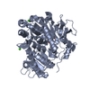
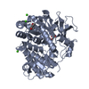
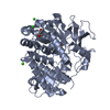
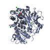
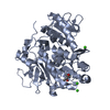
- PDB-9my7: Structure of the BasE mutant V336G, an NRPS adenylation domain in... -
+ Open data

- Basic information
| Entry | Database: PDB / ID: 9my7 | ||||||
|---|---|---|---|---|---|---|---|
| Title | Structure of the BasE mutant V336G, an NRPS adenylation domain in the acinetobactin biosynthetic pathway bound to 4-amino salicylic acid | ||||||
 Components Components | (2,3-dihydroxybenzoyl)adenylate synthase | ||||||
 Keywords Keywords | LIGASE / NRPS / Adenylation Domain / Nonribosomal peptide siderophore / acinetobactin / synthetase | ||||||
| Function / homology |  Function and homology information Function and homology information(2,3-dihydroxybenzoyl)adenylate synthase / 2,3-dihydroxybenzoate--[aryl-carrier protein] ligase / siderophore biosynthetic process / nucleotidyltransferase activity Similarity search - Function | ||||||
| Biological species |  Acinetobacter baumannii (bacteria) Acinetobacter baumannii (bacteria) | ||||||
| Method |  X-RAY DIFFRACTION / SYNCHROTRON / MOLECULAR REPLACEMENT / Resolution: 2.53 Å X-RAY DIFFRACTION / SYNCHROTRON / MOLECULAR REPLACEMENT / Resolution: 2.53 Å | ||||||
 Authors Authors | Ahmed, S.F. / Gulick, A.M. | ||||||
| Funding support |  United States, 1items United States, 1items
| ||||||
 Citation Citation | Journal: J.Biol.Chem. / Year: 2025 Title: The structural basis of substrate selectivity of the acinetobactin biosynthetic adenylation domain, BasE. Authors: Ahmed, S.F. / Gulick, A.M. | ||||||
| History |
|
- Structure visualization
| Structure viewer | Molecule: MolmilJmol/JSmol |
|---|
- Downloads & links
-Download
| PDBx/mmCIF format | 9my7.cif.gz | 423.1 KB | Display | PDBx/mmCIF format |
|---|---|---|---|---|
| PDB format | pdb9my7.ent.gz | 284.7 KB | Display | PDB format |
| PDBx/mmJSON format | 9my7.json.gz | Tree view | PDBx/mmJSON format | |
| Others |  Other downloads Other downloads |
-Validation report
| Summary document | 9my7_validation.pdf.gz | 909.9 KB | Display | wwPDB validaton report |
|---|---|---|---|---|
| Full document | 9my7_full_validation.pdf.gz | 926 KB | Display | |
| Data in XML | 9my7_validation.xml.gz | 39.6 KB | Display | |
| Data in CIF | 9my7_validation.cif.gz | 50.8 KB | Display | |
| Arichive directory | https://data.pdbj.org/pub/pdb/validation_reports/my/9my7ftp://data.pdbj.org/pub/pdb/validation_reports/my/9my7 | HTTPS FTP |
-Related structure data


-Links
 PDBj
PDBj
- Assembly
| Deposited unit | 
| ||||||||||||
|---|---|---|---|---|---|---|---|---|---|---|---|---|---|
| 1 | 
| ||||||||||||
| 2 | 
| ||||||||||||
| Unit cell |
|
-Components
| #1: Protein | Mass: 62905.395 Da / Num. of mol.: 2 / Mutation: P45L, V336G Source method: isolated from a genetically manipulated source Source: (gene. exp.) Acinetobacter baumannii (bacteria) / Strain: AB900 / Gene: entE, basE, ABR2091_2618, GSE42_14350, H0529_00955Production host: References: UniProt: A0A505MWF2, (2,3-dihydroxybenzoyl)adenylate synthase #2: Chemical | 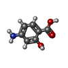  Mass: 153.135 Da / Num. of mol.: 2 / Source method: obtained synthetically / Formula: C7H7NO3 / Feature type: SUBJECT OF INVESTIGATION Mass: 153.135 Da / Num. of mol.: 2 / Source method: obtained synthetically / Formula: C7H7NO3 / Feature type: SUBJECT OF INVESTIGATION#3: Chemical |   Mass: 62.068 Da / Num. of mol.: 3 / Source method: obtained synthetically / Formula: C2H6O2 Mass: 62.068 Da / Num. of mol.: 3 / Source method: obtained synthetically / Formula: C2H6O2#4: Chemical | ChemComp-CA /   Mass: 40.078 Da / Num. of mol.: 9 / Source method: obtained synthetically / Formula: Ca Mass: 40.078 Da / Num. of mol.: 9 / Source method: obtained synthetically / Formula: Ca#5: Water | ChemComp-HOH / |  Mass: 18.015 Da / Num. of mol.: 153 / Source method: isolated from a natural source / Formula: H2O Mass: 18.015 Da / Num. of mol.: 153 / Source method: isolated from a natural source / Formula: H2OHas ligand of interest | Y | Has protein modification | N | Sequence details | This entry uses a Uniprot reference that is for a different strain of A. Baumannii. These sequence ...This entry uses a Uniprot reference that is for a different strain of A. Baumannii. These sequence discrepancies listed as "conflicts" are due to this strain difference. The wild-type sequence for the protein from this strain of A. Baumannii is found in Genbank entry WP_000744385.1. | |
|---|
-Experimental details
-Experiment
| Experiment | Method: X-RAY DIFFRACTION / Number of used crystals: 1 |
|---|
- Sample preparation
| Crystal | Density Matthews: 2.77 Å3/Da / Density % sol: 55.67 % |
|---|---|
| Crystal grow | Temperature: 287 K / Method: vapor diffusion, sitting drop / pH: 8.5 Details: 12% PEG 4000, 0.1 M Calcium chloride, 0.05 M TRIS HCl pH 8.5, 5mM 4-azidosalicylic acid |
-Data collection
| Diffraction | Mean temperature: 93 K / Serial crystal experiment: N |
|---|---|
| Diffraction source | Source: SYNCHROTRON / Site: SSRL / Beamline: BL12-2 / Wavelength: 0.97946 Å |
| Detector | Type: DECTRIS EIGER X 16M / Detector: PIXEL / Date: Jul 17, 2024 |
| Radiation | Protocol: SINGLE WAVELENGTH / Monochromatic (M) / Laue (L): M / Scattering type: x-ray |
| Radiation wavelength | Wavelength: 0.97946 Å / Relative weight: 1 |
| Reflection | Resolution: 2.53→66.17 Å / Num. obs: 47313 / % possible obs: 99.6 % / Redundancy: 3.9 % / Biso Wilson estimate: 50.66 Å2 / CC1/2: 0.996 / Rmerge(I) obs: 0.113 / Rpim(I) all: 0.063 / Net I/σ(I): 9.6 |
| Reflection shell | Resolution: 2.53→2.67 Å / Redundancy: 4 % / Rmerge(I) obs: 1.8 / Mean I/σ(I) obs: 2.9 / Num. unique obs: 6822 / CC1/2: 0.522 / Rpim(I) all: 1 / % possible all: 99.9 |
- Processing
| Software |
| |||||||||||||||||||||||||||||||||||||||||||||||||||||||||||||||||||||||||||||||||||||||||||||||||||||||||
|---|---|---|---|---|---|---|---|---|---|---|---|---|---|---|---|---|---|---|---|---|---|---|---|---|---|---|---|---|---|---|---|---|---|---|---|---|---|---|---|---|---|---|---|---|---|---|---|---|---|---|---|---|---|---|---|---|---|---|---|---|---|---|---|---|---|---|---|---|---|---|---|---|---|---|---|---|---|---|---|---|---|---|---|---|---|---|---|---|---|---|---|---|---|---|---|---|---|---|---|---|---|---|---|---|---|---|
| Refinement | Method to determine structure: MOLECULAR REPLACEMENT / Resolution: 2.53→66.17 Å / SU ML: 0.3527 / Cross valid method: FREE R-VALUE / σ(F): 1.33 / Phase error: 31.5133 Stereochemistry target values: GeoStd + Monomer Library + CDL v1.2
| |||||||||||||||||||||||||||||||||||||||||||||||||||||||||||||||||||||||||||||||||||||||||||||||||||||||||
| Solvent computation | Shrinkage radii: 0.9 Å / VDW probe radii: 1.1 Å / Solvent model: FLAT BULK SOLVENT MODEL | |||||||||||||||||||||||||||||||||||||||||||||||||||||||||||||||||||||||||||||||||||||||||||||||||||||||||
| Displacement parameters | Biso mean: 70.4 Å2 | |||||||||||||||||||||||||||||||||||||||||||||||||||||||||||||||||||||||||||||||||||||||||||||||||||||||||
| Refinement step | Cycle: LAST / Resolution: 2.53→66.17 Å
| |||||||||||||||||||||||||||||||||||||||||||||||||||||||||||||||||||||||||||||||||||||||||||||||||||||||||
| Refine LS restraints |
| |||||||||||||||||||||||||||||||||||||||||||||||||||||||||||||||||||||||||||||||||||||||||||||||||||||||||
| LS refinement shell |
| |||||||||||||||||||||||||||||||||||||||||||||||||||||||||||||||||||||||||||||||||||||||||||||||||||||||||
| Refinement TLS params. | Method: refined / Refine-ID: X-RAY DIFFRACTION
| |||||||||||||||||||||||||||||||||||||||||||||||||||||||||||||||||||||||||||||||||||||||||||||||||||||||||
| Refinement TLS group | Refine-ID: X-RAY DIFFRACTION / Auth seq-ID: 3 - 437 / Label seq-ID: 1 - 435
|
