Movie
Movie Controller


+ Open data

- Basic information
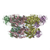
| Entry | Database: PDB / ID: 9mpu | ||||||||||||
|---|---|---|---|---|---|---|---|---|---|---|---|---|---|
| Title | Cryo-EM structure of p47 bound to VCP N-domain (with D1 domain) | ||||||||||||
 Components Components |
| ||||||||||||
 Keywords Keywords | HYDROLASE / double-ring hexameric complex / valosin containing protein / ATPase / VCP / mammalian / p97 / p47 / adapter / SHP / UBX | ||||||||||||
| Function / homology |  Function and homology information Function and homology informationnegative regulation of protein localization to centrosome / positive regulation of mitotic centrosome separation / nuclear membrane reassembly / flavin adenine dinucleotide catabolic process / VCP-NSFL1C complex / endoplasmic reticulum stress-induced pre-emptive quality control / endosome to lysosome transport via multivesicular body sorting pathway / Golgi stack / BAT3 complex binding / cellular response to arsenite ion ...negative regulation of protein localization to centrosome / positive regulation of mitotic centrosome separation / nuclear membrane reassembly / flavin adenine dinucleotide catabolic process / VCP-NSFL1C complex / endoplasmic reticulum stress-induced pre-emptive quality control / endosome to lysosome transport via multivesicular body sorting pathway / Golgi stack / BAT3 complex binding / cellular response to arsenite ion / cytoplasmic ubiquitin ligase complex / protein-DNA covalent cross-linking repair / Derlin-1 retrotranslocation complex / positive regulation of protein K63-linked deubiquitination / deubiquitinase activator activity / positive regulation of oxidative phosphorylation / cytoplasm protein quality control / aggresome assembly / ubiquitin-modified protein reader activity / regulation of protein localization to chromatin / cellular response to misfolded protein / mitotic spindle disassembly / VCP-NPL4-UFD1 AAA ATPase complex / positive regulation of mitochondrial membrane potential / vesicle-fusing ATPase / ATPase complex / K48-linked polyubiquitin modification-dependent protein binding / regulation of aerobic respiration / NAD+ metabolic process / retrograde protein transport, ER to cytosol / stress granule disassembly / Golgi organization / ubiquitin-specific protease binding / establishment of mitotic spindle orientation / regulation of synapse organization / ciliary transition zone / positive regulation of ATP biosynthetic process / intracellular membrane-bounded organelle / ubiquitin-like protein ligase binding / RHOH GTPase cycle / MHC class I protein binding / autophagosome maturation / negative regulation of hippo signaling / HSF1 activation / endoplasmic reticulum to Golgi vesicle-mediated transport / polyubiquitin modification-dependent protein binding / autophagosome assembly / ATP metabolic process / interstrand cross-link repair / Attachment and Entry / endoplasmic reticulum unfolded protein response / Protein methylation / ERAD pathway / ciliary tip / translesion synthesis / negative regulation of protein localization to chromatin / lipid droplet / viral genome replication / proteasome complex / Josephin domain DUBs / macroautophagy / proteasomal protein catabolic process / negative regulation of smoothened signaling pathway / ubiquitin binding / establishment of protein localization / N-glycan trimming in the ER and Calnexin/Calreticulin cycle / positive regulation of protein-containing complex assembly / ADP binding / Hh mutants are degraded by ERAD / Translesion Synthesis by POLH / positive regulation of non-canonical NF-kappaB signal transduction / Hedgehog ligand biogenesis / Defective CFTR causes cystic fibrosis / autophagy / ABC-family protein mediated transport / cytoplasmic stress granule / Aggrephagy / positive regulation of protein catabolic process / positive regulation of canonical Wnt signaling pathway / azurophil granule lumen / Ovarian tumor domain proteases / KEAP1-NFE2L2 pathway / double-strand break repair / positive regulation of proteasomal ubiquitin-dependent protein catabolic process / cellular response to heat / chromosome / E3 ubiquitin ligases ubiquitinate target proteins / site of double-strand break / Neddylation / secretory granule lumen / protein phosphatase binding / regulation of apoptotic process / ficolin-1-rich granule lumen / ubiquitin-dependent protein catabolic process / membrane fusion / Attachment and Entry / proteasome-mediated ubiquitin-dependent protein catabolic process / ciliary basal body / protein ubiquitination / protein domain specific binding Similarity search - Function | ||||||||||||
| Biological species |  Homo sapiens (human) Homo sapiens (human) | ||||||||||||
| Method | ELECTRON MICROSCOPY / single particle reconstruction / cryo EM / Resolution: 4 Å | ||||||||||||
 Authors Authors | Shah, B. / Hunkeler, M. / Buhrlage, S.J. / Fischer, E.F. | ||||||||||||
| Funding support |  United States, 3items United States, 3items
| ||||||||||||
 Citation Citation | Journal: Nat Commun / Year: 2025 Title: Structural basis of VCP-VCPIP1-p47 ternary complex in Golgi maintenance. Authors: Binita Shah / Moritz Hunkeler / Ariana Bratt / Hong Yue / Isabella Jaen Maisonet / Eric S Fischer / Sara J Buhrlage / Abstract: VCP/p97 regulates a wide range of cellular processes, including post-mitotic Golgi reassembly. In this context, VCP is assisted by p47, an adapter protein, and VCPIP1, a deubiquitylase (DUB). ...VCP/p97 regulates a wide range of cellular processes, including post-mitotic Golgi reassembly. In this context, VCP is assisted by p47, an adapter protein, and VCPIP1, a deubiquitylase (DUB). However, how they organize into a functional ternary complex to promote Golgi assembly remains unknown. Here, we use cryo-EM to characterize both VCP-VCPIP1 and VCP-VCPIP1-p47 complexes. We show that VCPIP1 engages VCP through two interfaces: one involving the N-domain of VCP and the UBX domain of VCPIP1, and the other involving the VCP D2 domains and a region of VCPIP1 we refer to as VCPID. The p47 UBX domain competitively binds to the VCP N-domain, while not affecting VCPID binding. We show that VCPID is critical for VCP-mediated enhancement of DUB activity and proper Golgi assembly. The ternary structure along with biochemical and cellular data provides new insights into the complex interplay of VCP with its co-factors. | ||||||||||||
| History |
|
- Structure visualization
| Structure viewer | Molecule: MolmilJmol/JSmol |
|---|
- Downloads & links
-Download
| PDBx/mmCIF format | 9mpu.cif.gz | 244 KB | Display | PDBx/mmCIF format |
|---|---|---|---|---|
| PDB format | pdb9mpu.ent.gz | 157.7 KB | Display | PDB format |
| PDBx/mmJSON format | 9mpu.json.gz | Tree view | PDBx/mmJSON format | |
| Others |  Other downloads Other downloads |
-Validation report
| Arichive directory | https://data.pdbj.org/pub/pdb/validation_reports/mp/9mpuftp://data.pdbj.org/pub/pdb/validation_reports/mp/9mpu | HTTPS FTP |
|---|
-Related structure data
| Related structure data |  48505MC  9mpqC  9mprC  9mpsC  9mptC  9mpvC C: citing same article ( M: map data used to model this data |
|---|---|
| Similar structure data |


-Links
 PDBj
PDBj


- Assembly
| Deposited unit | 
|
|---|---|
| 1 |
|
-Components
| #1: Protein | Mass: 43535.906 Da / Num. of mol.: 1 Source method: isolated from a genetically manipulated source Details: N-term His tagged p47 / Source: (gene. exp.) Homo sapiens (human) / Gene: NSFL1C, UBXN2C / Production host:  |
|---|---|
| #2: Protein | Mass: 92022.539 Da / Num. of mol.: 1 Source method: isolated from a genetically manipulated source Details: N-term FLAG-tagged VCP / Source: (gene. exp.) Homo sapiens (human) / Gene: VCP, HEL-220, HEL-S-70 / Cell (production host): Expi293 / Production host: Homo sapiens (human) / References: UniProt: P55072, vesicle-fusing ATPase |
| Has protein modification | N |
-Experimental details
-Experiment
| Experiment | Method: ELECTRON MICROSCOPY |
|---|---|
| EM experiment | Aggregation state: PARTICLE / 3D reconstruction method: single particle reconstruction |
- Sample preparation
| Component |
| ||||||||||||||||||||||||||||
|---|---|---|---|---|---|---|---|---|---|---|---|---|---|---|---|---|---|---|---|---|---|---|---|---|---|---|---|---|---|
| Molecular weight |
| ||||||||||||||||||||||||||||
| Source (natural) |
| ||||||||||||||||||||||||||||
| Source (recombinant) |
| ||||||||||||||||||||||||||||
| Buffer solution | pH: 7.4 | ||||||||||||||||||||||||||||
| Buffer component |
| ||||||||||||||||||||||||||||
| Specimen | Conc.: 2 mg/ml / Embedding applied: NO / Shadowing applied: NO / Staining applied: NO / Vitrification applied: YES / Details: Final concentration of 0.2 mM CHAPSO | ||||||||||||||||||||||||||||
| Specimen support | Grid material: COPPER / Grid mesh size: 300 divisions/in. / Grid type: Quantifoil R1.2/1.3 | ||||||||||||||||||||||||||||
| Vitrification | Instrument: LEICA EM GP / Cryogen name: ETHANE / Humidity: 90 % / Chamber temperature: 283.15 K |
- Electron microscopy imaging
| Experimental equipment |  Model: Titan Krios / Image courtesy: FEI Company |
|---|---|
| Microscopy | Model: TFS KRIOS |
| Electron gun | Electron source:  FIELD EMISSION GUN / Accelerating voltage: 300 kV / Illumination mode: FLOOD BEAM FIELD EMISSION GUN / Accelerating voltage: 300 kV / Illumination mode: FLOOD BEAM |
| Electron lens | Mode: BRIGHT FIELD / Nominal magnification: 165000 X / Nominal defocus max: 2200 nm / Nominal defocus min: 800 nm / Cs: 2.7 mm / Alignment procedure: COMA FREE |
| Specimen holder | Cryogen: NITROGEN / Specimen holder model: FEI TITAN KRIOS AUTOGRID HOLDER |
| Image recording | Average exposure time: 2.87 sec. / Electron dose: 49.22 e/Å2 / Film or detector model: FEI FALCON IV (4k x 4k) / Num. of grids imaged: 1 / Num. of real images: 12261 |
| EM imaging optics | Energyfilter slit width: 10 eV |
- Processing
| EM software |
| ||||||||||||||||||||||||||||||||||||||||||||||||||
|---|---|---|---|---|---|---|---|---|---|---|---|---|---|---|---|---|---|---|---|---|---|---|---|---|---|---|---|---|---|---|---|---|---|---|---|---|---|---|---|---|---|---|---|---|---|---|---|---|---|---|---|
| CTF correction | Type: PHASE FLIPPING AND AMPLITUDE CORRECTION | ||||||||||||||||||||||||||||||||||||||||||||||||||
| Particle selection | Num. of particles selected: 2003371 | ||||||||||||||||||||||||||||||||||||||||||||||||||
| 3D reconstruction | Resolution: 4 Å / Resolution method: FSC 0.143 CUT-OFF / Num. of particles: 54855 / Algorithm: FOURIER SPACE / Symmetry type: POINT | ||||||||||||||||||||||||||||||||||||||||||||||||||
| Atomic model building | Space: REAL | ||||||||||||||||||||||||||||||||||||||||||||||||||
| Atomic model building | PDB-ID: 5FTK Accession code: 5FTK / Source name: PDB / Type: experimental model | ||||||||||||||||||||||||||||||||||||||||||||||||||
| Refinement | Cross valid method: NONE Stereochemistry target values: GeoStd + Monomer Library + CDL v1.2 | ||||||||||||||||||||||||||||||||||||||||||||||||||
| Displacement parameters | Biso mean: 145.09 Å2 | ||||||||||||||||||||||||||||||||||||||||||||||||||
| Refine LS restraints |
|