Movie
Movie Controller

[English] 日本語
 Yorodumi
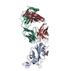
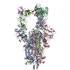
- PDB-9ml5: Structure of the SARS-CoV-2 Spike 6P in complex with the rabbit M... -
+ Open data

- Basic information
| Entry | Database: PDB / ID: 9ml5 | |||||||||
|---|---|---|---|---|---|---|---|---|---|---|
| Title | Structure of the SARS-CoV-2 Spike 6P in complex with the rabbit M8b-B8 Fab | |||||||||
 Components Components |
| |||||||||
 Keywords Keywords | VIRAL PROTEIN/IMMUNE SYSTEM / immune system / neutralizing antibody / IMMUNE SYSTEM-VIRAL PROTEIN complex / VIRAL PROTEIN-IMMUNE SYSTEM complex | |||||||||
| Function / homology |  Function and homology information Function and homology informationsymbiont-mediated disruption of host tissue / Maturation of spike protein / Translation of Structural Proteins / Virion Assembly and Release / host cell surface / host extracellular region / symbiont-mediated-mediated suppression of host tetherin activity / Induction of Cell-Cell Fusion / structural constituent of virion / positive regulation of viral entry into host cell ...symbiont-mediated disruption of host tissue / Maturation of spike protein / Translation of Structural Proteins / Virion Assembly and Release / host cell surface / host extracellular region / symbiont-mediated-mediated suppression of host tetherin activity / Induction of Cell-Cell Fusion / structural constituent of virion / positive regulation of viral entry into host cell / membrane fusion / host cell endoplasmic reticulum-Golgi intermediate compartment membrane / Attachment and Entry / entry receptor-mediated virion attachment to host cell / receptor-mediated virion attachment to host cell / host cell surface receptor binding / symbiont-mediated suppression of host innate immune response / endocytosis involved in viral entry into host cell / receptor ligand activity / fusion of virus membrane with host plasma membrane / fusion of virus membrane with host endosome membrane / viral envelope / symbiont entry into host cell / virion attachment to host cell / host cell plasma membrane / SARS-CoV-2 activates/modulates innate and adaptive immune responses / virion membrane / membrane / identical protein binding / plasma membrane Similarity search - Function | |||||||||
| Biological species |   Severe acute respiratory syndrome coronavirus 2 Severe acute respiratory syndrome coronavirus 2 | |||||||||
| Method | ELECTRON MICROSCOPY / single particle reconstruction / cryo EM / Resolution: 3.4 Å | |||||||||
 Authors Authors | Fan, C. / Bjorkman, P.J. | |||||||||
| Funding support |  United States, 2items United States, 2items
| |||||||||
 Citation Citation | Journal: Proc Natl Acad Sci U S A / Year: 2025 Title: Cross-reactive sarbecovirus antibodies induced by mosaic RBD nanoparticles. Authors: Chengcheng Fan / Jennifer R Keeffe / Kathryn E Malecek / Alexander A Cohen / Anthony P West / Viren A Baharani / Annie V Rorick / Han Gao / Priyanthi N P Gnanapragasam / Semi Rho / Jaasiel ...Authors: Chengcheng Fan / Jennifer R Keeffe / Kathryn E Malecek / Alexander A Cohen / Anthony P West / Viren A Baharani / Annie V Rorick / Han Gao / Priyanthi N P Gnanapragasam / Semi Rho / Jaasiel Alvarez / Luisa N Segovia / Theodora Hatziioannou / Paul D Bieniasz / Pamela J Bjorkman / Abstract: Broad immune responses are needed to mitigate viral evolution and escape. To induce antibodies against conserved receptor-binding domain (RBD) regions of SARS-like betacoronavirus (sarbecovirus) ...Broad immune responses are needed to mitigate viral evolution and escape. To induce antibodies against conserved receptor-binding domain (RBD) regions of SARS-like betacoronavirus (sarbecovirus) spike proteins that recognize SARS-CoV-2 variants of concern and zoonotic sarbecoviruses, we developed mosaic-8b RBD nanoparticles presenting eight sarbecovirus RBDs arranged randomly on a 60-mer nanoparticle. Mosaic-8b immunizations protected animals from challenges from viruses whose RBDs were matched or mismatched to those on nanoparticles. Here, we describe neutralizing mAbs isolated from mosaic-8b-immunized rabbits, some on par with Pemgarda, the only currently FDA-approved therapeutic mAb. Deep mutational scanning, in vitro selection of spike resistance mutations, and single-particle cryo-electron microscopy structures of spike-antibody complexes demonstrated targeting of conserved RBD epitopes. Rabbit mAbs included critical D-gene segment RBD-recognizing features in common with human anti-RBD mAbs, despite rabbit genomes lacking an equivalent human D-gene segment, thus demonstrating that the immune systems of humans and other mammals can utilize different antibody gene segments to arrive at similar modes of antigen recognition. These results suggest that animal models can be used to elicit anti-RBD mAbs with similar properties to those raised in humans, which can then be humanized for therapeutic use, and that mosaic RBD nanoparticle immunization coupled with multiplexed screening represents an efficient way to generate and select broadly cross-reactive therapeutic pan-sarbecovirus and pan-SARS-CoV-2 variant mAbs. | |||||||||
| History |
|
- Structure visualization
| Structure viewer | Molecule: MolmilJmol/JSmol |
|---|
- Downloads & links
-Download
| PDBx/mmCIF format | 9ml5.cif.gz | 872.5 KB | Display | PDBx/mmCIF format |
|---|---|---|---|---|
| PDB format | pdb9ml5.ent.gz | 570.2 KB | Display | PDB format |
| PDBx/mmJSON format | 9ml5.json.gz | Tree view | PDBx/mmJSON format | |
| Others |  Other downloads Other downloads |
-Validation report
| Arichive directory | https://data.pdbj.org/pub/pdb/validation_reports/ml/9ml5ftp://data.pdbj.org/pub/pdb/validation_reports/ml/9ml5 | HTTPS FTP |
|---|
-Related structure data
| Related structure data |  48348MC 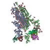 9ml4C 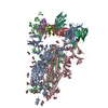 9ml6C 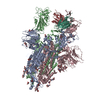 9ml7C  9ml8C  9ml9C M: map data used to model this data C: citing same article ( |
|---|---|
| Similar structure data |
-Links
 PDBj
PDBj


- Assembly
| Deposited unit | 
|
|---|---|
| 1 |
|
-Components
| #1: Protein | Mass: 139344.438 Da / Num. of mol.: 3 / Mutation: F817P, A892P, A899P, A942P, K986P, V987P Source method: isolated from a genetically manipulated source Source: (gene. exp.) Severe acute respiratory syndrome coronavirus 2Gene: S, 2 / Production host:  Homo sapiens (human) / References: UniProt: P0DTC2 Homo sapiens (human) / References: UniProt: P0DTC2#2: Protein | Mass: 23555.324 Da / Num. of mol.: 2 Source method: isolated from a genetically manipulated source Source: (gene. exp.) Homo sapiens (human)#3: Antibody | Mass: 23683.234 Da / Num. of mol.: 2 Source method: isolated from a genetically manipulated source Source: (gene. exp.) Homo sapiens (human)#4: Polysaccharide | 2-acetamido-2-deoxy-beta-D-glucopyranose-(1-4)-2-acetamido-2-deoxy-beta-D-glucopyranose Source method: isolated from a genetically manipulated source #5: Sugar | ChemComp-NAG /   Type: D-saccharide, beta linking / Mass: 221.208 Da / Num. of mol.: 31 / Source method: obtained synthetically / Formula: C8H15NO6 / Feature type: SUBJECT OF INVESTIGATION Type: D-saccharide, beta linking / Mass: 221.208 Da / Num. of mol.: 31 / Source method: obtained synthetically / Formula: C8H15NO6 / Feature type: SUBJECT OF INVESTIGATIONHas ligand of interest | Y | Has protein modification | Y | |
|---|
-Experimental details
-Experiment
| Experiment | Method: ELECTRON MICROSCOPY |
|---|---|
| EM experiment | Aggregation state: PARTICLE / 3D reconstruction method: single particle reconstruction |
- Sample preparation
| Component |
| ||||||||||||||||||||||||||||
|---|---|---|---|---|---|---|---|---|---|---|---|---|---|---|---|---|---|---|---|---|---|---|---|---|---|---|---|---|---|
| Molecular weight |
| ||||||||||||||||||||||||||||
| Source (natural) |
| ||||||||||||||||||||||||||||
| Source (recombinant) |
| ||||||||||||||||||||||||||||
| Buffer solution | pH: 8 | ||||||||||||||||||||||||||||
| Buffer component |
| ||||||||||||||||||||||||||||
| Specimen | Conc.: 2 mg/ml / Embedding applied: NO / Shadowing applied: NO / Staining applied: NO / Vitrification applied: YES | ||||||||||||||||||||||||||||
| Specimen support | Grid type: Quantifoil R1.2/1.3 | ||||||||||||||||||||||||||||
| Vitrification | Instrument: FEI VITROBOT MARK IV / Cryogen name: ETHANE / Humidity: 100 % / Chamber temperature: 293 K |
- Electron microscopy imaging
| Experimental equipment |  Model: Talos Arctica / Image courtesy: FEI Company |
|---|---|
| Microscopy | Model: FEI TALOS ARCTICA |
| Electron gun | Electron source:  FIELD EMISSION GUN / Accelerating voltage: 200 kV / Illumination mode: FLOOD BEAM FIELD EMISSION GUN / Accelerating voltage: 200 kV / Illumination mode: FLOOD BEAM |
| Electron lens | Mode: BRIGHT FIELD / Nominal magnification: 45000 X / Nominal defocus max: 3000 nm / Nominal defocus min: 1000 nm / Cs: 2.7 mm / Alignment procedure: COMA FREE |
| Specimen holder | Cryogen: NITROGEN / Specimen holder model: FEI TITAN KRIOS AUTOGRID HOLDER |
| Image recording | Average exposure time: 1.5 sec. / Electron dose: 60 e/Å2 / Film or detector model: GATAN K3 BIOQUANTUM (6k x 4k) / Num. of real images: 2634 |
- Processing
| EM software |
| ||||||||||||||||||||||||||||||||||||||||
|---|---|---|---|---|---|---|---|---|---|---|---|---|---|---|---|---|---|---|---|---|---|---|---|---|---|---|---|---|---|---|---|---|---|---|---|---|---|---|---|---|---|
| CTF correction | Type: PHASE FLIPPING AND AMPLITUDE CORRECTION | ||||||||||||||||||||||||||||||||||||||||
| Particle selection | Num. of particles selected: 576218 | ||||||||||||||||||||||||||||||||||||||||
| Symmetry | Point symmetry: C1 (asymmetric) | ||||||||||||||||||||||||||||||||||||||||
| 3D reconstruction | Resolution: 3.4 Å / Resolution method: FSC 0.143 CUT-OFF / Num. of particles: 148375 / Symmetry type: POINT | ||||||||||||||||||||||||||||||||||||||||
| Atomic model building | B value: 90 / Protocol: RIGID BODY FIT / Space: REAL | ||||||||||||||||||||||||||||||||||||||||
| Atomic model building |
| ||||||||||||||||||||||||||||||||||||||||
| Refinement | Cross valid method: NONE Stereochemistry target values: GeoStd + Monomer Library + CDL v1.2 | ||||||||||||||||||||||||||||||||||||||||
| Displacement parameters | Biso mean: 124.91 Å2 | ||||||||||||||||||||||||||||||||||||||||
| Refine LS restraints |
|