National Institutes of Health/National Institute Of Allergy and Infectious Diseases (NIH/NIAID)
P01-AI165075
United States
Bill & Melinda Gates Foundation
United States
Citation
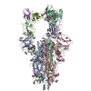
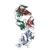
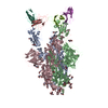
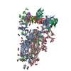
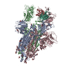
Journal: Proc Natl Acad Sci U S A / Year: 2025 Title: Cross-reactive sarbecovirus antibodies induced by mosaic RBD nanoparticles. Authors: Chengcheng Fan / Jennifer R Keeffe / Kathryn E Malecek / Alexander A Cohen / Anthony P West / Viren A Baharani / Annie V Rorick / Han Gao / Priyanthi N P Gnanapragasam / Semi Rho / Jaasiel ...Authors: Chengcheng Fan / Jennifer R Keeffe / Kathryn E Malecek / Alexander A Cohen / Anthony P West / Viren A Baharani / Annie V Rorick / Han Gao / Priyanthi N P Gnanapragasam / Semi Rho / Jaasiel Alvarez / Luisa N Segovia / Theodora Hatziioannou / Paul D Bieniasz / Pamela J Bjorkman / Abstract: Broad immune responses are needed to mitigate viral evolution and escape. To induce antibodies against conserved receptor-binding domain (RBD) regions of SARS-like betacoronavirus (sarbecovirus) ...Broad immune responses are needed to mitigate viral evolution and escape. To induce antibodies against conserved receptor-binding domain (RBD) regions of SARS-like betacoronavirus (sarbecovirus) spike proteins that recognize SARS-CoV-2 variants of concern and zoonotic sarbecoviruses, we developed mosaic-8b RBD nanoparticles presenting eight sarbecovirus RBDs arranged randomly on a 60-mer nanoparticle. Mosaic-8b immunizations protected animals from challenges from viruses whose RBDs were matched or mismatched to those on nanoparticles. Here, we describe neutralizing mAbs isolated from mosaic-8b-immunized rabbits, some on par with Pemgarda, the only currently FDA-approved therapeutic mAb. Deep mutational scanning, in vitro selection of spike resistance mutations, and single-particle cryo-electron microscopy structures of spike-antibody complexes demonstrated targeting of conserved RBD epitopes. Rabbit mAbs included critical D-gene segment RBD-recognizing features in common with human anti-RBD mAbs, despite rabbit genomes lacking an equivalent human D-gene segment, thus demonstrating that the immune systems of humans and other mammals can utilize different antibody gene segments to arrive at similar modes of antigen recognition. These results suggest that animal models can be used to elicit anti-RBD mAbs with similar properties to those raised in humans, which can then be humanized for therapeutic use, and that mosaic RBD nanoparticle immunization coupled with multiplexed screening represents an efficient way to generate and select broadly cross-reactive therapeutic pan-sarbecovirus and pan-SARS-CoV-2 variant mAbs.
In the structure databanks used in Yorodumi, some data are registered as the other names, "COVID-19 virus" and "2019-nCoV". Here are the details of the virus and the list of structure data.
Jan 31, 2019. EMDB accession codes are about to change! (news from PDBe EMDB page)
EMDB accession codes are about to change! (news from PDBe EMDB page)
The allocation of 4 digits for EMDB accession codes will soon come to an end. Whilst these codes will remain in use, new EMDB accession codes will include an additional digit and will expand incrementally as the available range of codes is exhausted. The current 4-digit format prefixed with “EMD-” (i.e. EMD-XXXX) will advance to a 5-digit format (i.e. EMD-XXXXX), and so on. It is currently estimated that the 4-digit codes will be depleted around Spring 2019, at which point the 5-digit format will come into force.
The EM Navigator/Yorodumi systems omit the EMD- prefix.
Related info.:Q: What is EMD? / ID/Accession-code notation in Yorodumi/EM Navigator
Yorodumi is a browser for structure data from EMDB, PDB, SASBDB, etc.
This page is also the successor to EM Navigator detail page, and also detail information page/front-end page for Omokage search.
The word "yorodu" (or yorozu) is an old Japanese word meaning "ten thousand". "mi" (miru) is to see.
Related info.:EMDB / PDB / SASBDB / Comparison of 3 databanks / Yorodumi Search / Aug 31, 2016. New EM Navigator & Yorodumi / Yorodumi Papers / Jmol/JSmol / Function and homology information / Changes in new EM Navigator and Yorodumi
 Movie
Movie Controller

 Yorodumi
 Open data
 Basic information
 Map data
 Sample
 Keywords
 Function and homology information
 Severe acute respiratory syndrome coronavirus 2 /
 Authors
 United States, 2 items
  Citation
 Structure visualization
 Downloads & links
 emd_48347.png
 http://ftp.pdbj.org/pub/emdb/structures/EMD-48347
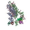


 Z (Sec.)
 Y (Row.)
 X (Col.)


 Sample components
 Homo sapiens (human)
 Processing
 Electron microscopy
 FIELD EMISSION GUN