 Movie
Movie Controller

[English] 日本語
 Yorodumi
- PDB-9mhz: Cryo-EM structure of S. aureus TarGH in complex with Targocil-II ... -
+ Open data

- Basic information
| Entry | Database: PDB / ID: 9mhz | ||||||
|---|---|---|---|---|---|---|---|
| Title | Cryo-EM structure of S. aureus TarGH in complex with Targocil-II and ATP-gamma-S in a catalytically incompetent conformation | ||||||
 Components Components |
| ||||||
 Keywords Keywords | MEMBRANE PROTEIN / ABC transporter / teichoic acid / bacteria | ||||||
| Function / homology |  Function and homology information Function and homology informationABC-type teichoic-acid transporter / ABC-type teichoic acid transporter activity / lipopolysaccharide transport / ABC-type transporter activity / ATP hydrolysis activity / ATP binding / plasma membrane Similarity search - Function | ||||||
| Biological species |   Staphylococcus aureus (bacteria) Staphylococcus aureus (bacteria) | ||||||
| Method | ELECTRON MICROSCOPY / single particle reconstruction / cryo EM / Resolution: 2.7 Å | ||||||
 Authors Authors | Peters, S.C. / Worrall, L.J. / Strynadka, N.C.J. | ||||||
| Funding support |  Canada, 1items Canada, 1items
| ||||||
 Citation Citation | Journal: Nat Commun / Year: 2025 Title: Cryo-EM analyses unveil details of mechanism and targocil-II mediated inhibition of S. aureus WTA transporter TarGH. Authors: Franco K K Li / Shaun C Peters / Liam J Worrall / Tianjun Sun / Jinhong Hu / Marija Vuckovic / Maya Farha / Armando Palacios / Nathanael A Caveney / Eric D Brown / Natalie C J Strynadka / Abstract: Wall teichoic acid (WTA) is a polyol phosphate polymer that covalently decorates peptidoglycan of gram-positive bacteria, including Staphylococcus aureus. Central to WTA biosynthesis is flipping of ...Wall teichoic acid (WTA) is a polyol phosphate polymer that covalently decorates peptidoglycan of gram-positive bacteria, including Staphylococcus aureus. Central to WTA biosynthesis is flipping of lipid-linked precursors across the cell membrane by TarGH, a type V ABC transporter. Here, we present cryo-EM structures of S. aureus TarGH in the presence of targocil-II, a promising small-molecule lead with β-lactam antibiotic synergistic action. Targocil-II binds to the extracellular dimerisation interface of TarG, we suggest mimicking flipped but not yet released substrate. In absence of targocil-II and in complex with ATP analogue ATPγS, determined at 2.3 Å resolution, the ATPase active site is allosterically inhibited. This is due to a so far undescribed D-loop conformation, potentially minimizing spurious ATP hydrolysis in the absence of substrate. Targocil-II binding comparatively causes local and remote conformational changes through to the TarH active site, with the D-loop now optimal for ATP hydrolysis. These structures suggest an ability to modulate ATP hydrolysis in a WTA substrate dependent manner and a jammed ATPase cycle as the basis of the observed inhibition by targocil-II. The molecular insights provide an unprecedented basis for development of TarGH targeted therapeutics for treatment of multidrug-resistant S. aureus and other gram-positive bacterial infections. | ||||||
| History |
|
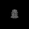
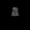
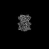
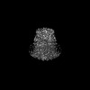
- Structure visualization
| Structure viewer | Molecule: MolmilJmol/JSmol |
|---|
- Downloads & links
-Download
| PDBx/mmCIF format | 9mhz.cif.gz | 232 KB | Display | PDBx/mmCIF format |
|---|---|---|---|---|
| PDB format | pdb9mhz.ent.gz | Display | PDB format | |
| PDBx/mmJSON format | 9mhz.json.gz | Tree view | PDBx/mmJSON format | |
| Others |  Other downloads Other downloads |
-Validation report
| Summary document | 9mhz_validation.pdf.gz | 1.7 MB | Display | wwPDB validaton report |
|---|---|---|---|---|
| Full document | 9mhz_full_validation.pdf.gz | 1.7 MB | Display | |
| Data in XML | 9mhz_validation.xml.gz | 42.9 KB | Display | |
| Data in CIF | 9mhz_validation.cif.gz | 61.6 KB | Display | |
| Arichive directory | https://data.pdbj.org/pub/pdb/validation_reports/mh/9mhzftp://data.pdbj.org/pub/pdb/validation_reports/mh/9mhz | HTTPS FTP |
-Related structure data
| Related structure data |  48282MC 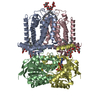 9cflC  9cfpC 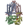 9mhdC 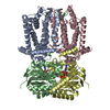 9mhuC C: citing same article ( M: map data used to model this data |
|---|---|
| Similar structure data |
-Links
 PDBj
PDBj


- Assembly
| Deposited unit | 
|
|---|---|
| 1 |
|
-Components
-Protein , 2 types, 4 molecules ACBD
| #1: Protein | Mass: 34240.527 Da / Num. of mol.: 2 Source method: isolated from a genetically manipulated source Source: (gene. exp.) Staphylococcus aureus (bacteria) / Gene: tagG, SAUSA300_0625 / Production host: Lactococcus lactis (lactic acid bacteria) / References: UniProt: A0A0H2XIF1#2: Protein | Mass: 29806.553 Da / Num. of mol.: 2 Source method: isolated from a genetically manipulated source Source: (gene. exp.) Staphylococcus aureus (bacteria) / Gene: tagH, SAUSA300_0624 / Production host: Lactococcus lactis (lactic acid bacteria)References: UniProt: Q2FJ01, ABC-type teichoic-acid transporter |
|---|
-Non-polymers , 5 types, 16 molecules 


| #3: Chemical |  Mass: 1005.188 Da / Num. of mol.: 2 / Source method: obtained synthetically / Formula: C47H88O22 / Feature type: SUBJECT OF INVESTIGATION Mass: 1005.188 Da / Num. of mol.: 2 / Source method: obtained synthetically / Formula: C47H88O22 / Feature type: SUBJECT OF INVESTIGATION#4: Chemical | Mass: 479.909 Da / Num. of mol.: 2 / Source method: obtained synthetically / Formula: C26H22ClNO6 / Feature type: SUBJECT OF INVESTIGATION #5: Chemical |  Mass: 523.247 Da / Num. of mol.: 2 / Source method: isolated from a natural source / Formula: C10H16N5O12P3S / Feature type: SUBJECT OF INVESTIGATION / Comment: ATP-gamma-S, energy-carrying molecule analogue*YM Mass: 523.247 Da / Num. of mol.: 2 / Source method: isolated from a natural source / Formula: C10H16N5O12P3S / Feature type: SUBJECT OF INVESTIGATION / Comment: ATP-gamma-S, energy-carrying molecule analogue*YM#6: Chemical |  Mass: 24.305 Da / Num. of mol.: 2 / Source method: obtained synthetically / Formula: Mg / Feature type: SUBJECT OF INVESTIGATION Mass: 24.305 Da / Num. of mol.: 2 / Source method: obtained synthetically / Formula: Mg / Feature type: SUBJECT OF INVESTIGATION#7: Water | ChemComp-HOH / | Mass: 18.015 Da / Num. of mol.: 8 / Source method: isolated from a natural source / Formula: H2O |
|---|
-Details
| Has ligand of interest | Y |
|---|---|
| Has protein modification | N |
-Experimental details
-Experiment
| Experiment | Method: ELECTRON MICROSCOPY |
|---|---|
| EM experiment | Aggregation state: PARTICLE / 3D reconstruction method: single particle reconstruction |
- Sample preparation
| Component | Name: TarGH / Type: COMPLEX / Details: Hetero tetramer TarG2H2 / Entity ID: #1-#2 / Source: RECOMBINANT |
|---|---|
| Molecular weight | Experimental value: NO |
| Source (natural) | Organism: Staphylococcus aureus (bacteria) |
| Source (recombinant) | Organism: Lactococcus lactis (lactic acid bacteria) |
| Buffer solution | pH: 8 |
| Specimen | Embedding applied: NO / Shadowing applied: NO / Staining applied: NO / Vitrification applied: YES |
| Vitrification | Cryogen name: ETHANE |
- Electron microscopy imaging
| Experimental equipment |  Model: Titan Krios / Image courtesy: FEI Company |
|---|---|
| Microscopy | Model: TFS KRIOS |
| Electron gun | Electron source:  FIELD EMISSION GUN / Accelerating voltage: 300 kV / Illumination mode: FLOOD BEAM FIELD EMISSION GUN / Accelerating voltage: 300 kV / Illumination mode: FLOOD BEAM |
| Electron lens | Mode: BRIGHT FIELD / Nominal defocus max: 3000 nm / Nominal defocus min: 500 nm |
| Image recording | Electron dose: 50 e/Å2 / Film or detector model: TFS FALCON 4i (4k x 4k) |
- Processing
| EM software | Name: PHENIX / Version: 1.20.1_4487: / Category: model refinement | ||||||||||||||||||||||||
|---|---|---|---|---|---|---|---|---|---|---|---|---|---|---|---|---|---|---|---|---|---|---|---|---|---|
| CTF correction | Type: PHASE FLIPPING AND AMPLITUDE CORRECTION | ||||||||||||||||||||||||
| 3D reconstruction | Resolution: 2.7 Å / Resolution method: FSC 0.143 CUT-OFF / Num. of particles: 121556 / Symmetry type: POINT | ||||||||||||||||||||||||
| Refine LS restraints |
|
