 Movie
Movie Controller

[English] 日本語
 Yorodumi
- PDB-9m2h: Structure of the auxin importer AUX1 in Arabidopsis thaliana in t... -
+ Open data

- Basic information
| Entry | Database: PDB / ID: 9m2h | |||||||||||||||||||||||||||
|---|---|---|---|---|---|---|---|---|---|---|---|---|---|---|---|---|---|---|---|---|---|---|---|---|---|---|---|---|
| Title | Structure of the auxin importer AUX1 in Arabidopsis thaliana in the CHPAA-bound state | |||||||||||||||||||||||||||
 Components Components | Auxin transporter protein 1 | |||||||||||||||||||||||||||
 Keywords Keywords | MEMBRANE PROTEIN / Auxin / importer / AUX1 | |||||||||||||||||||||||||||
| Function / homology |  Function and homology information Function and homology informationroot hair cell differentiation / auxin binding / root cap development / somatic embryogenesis / lateral root formation / auxin influx transmembrane transporter activity / positive gravitropism / establishment of planar polarity / auxin polar transport / amino acid transmembrane transport ...root hair cell differentiation / auxin binding / root cap development / somatic embryogenesis / lateral root formation / auxin influx transmembrane transporter activity / positive gravitropism / establishment of planar polarity / auxin polar transport / amino acid transmembrane transport / auxin-activated signaling pathway / response to nematode / symporter activity / amino acid transmembrane transporter activity / endosome / cell surface / Golgi apparatus / plasma membrane Similarity search - Function | |||||||||||||||||||||||||||
| Biological species |  | |||||||||||||||||||||||||||
| Method | ELECTRON MICROSCOPY / single particle reconstruction / cryo EM / Resolution: 3.4 Å | |||||||||||||||||||||||||||
 Authors Authors | Sun, L. / Liu, X. / Wei, H. / Yang, Z. | |||||||||||||||||||||||||||
| Funding support |  China, 3items China, 3items
| |||||||||||||||||||||||||||
 Citation Citation | Journal: To Be Published Title: Structure of the auxin importer AUX1 in Arabidopsis thaliana in the CHPAA-bound state Authors: Sun, L. / Liu, X. / Wei, H. / Yang, Z. | |||||||||||||||||||||||||||
| History |
|
- Structure visualization
| Structure viewer | Molecule: MolmilJmol/JSmol |
|---|
- Downloads & links
-Download
| PDBx/mmCIF format | 9m2h.cif.gz | 87.6 KB | Display | PDBx/mmCIF format |
|---|---|---|---|---|
| PDB format | pdb9m2h.ent.gz | 65 KB | Display | PDB format |
| PDBx/mmJSON format | 9m2h.json.gz | Tree view | PDBx/mmJSON format | |
| Others |  Other downloads Other downloads |
-Validation report
| Arichive directory | https://data.pdbj.org/pub/pdb/validation_reports/m2/9m2hftp://data.pdbj.org/pub/pdb/validation_reports/m2/9m2h | HTTPS FTP |
|---|
-Related structure data
| Related structure data |  63585MC M: map data used to model this data C: citing same article ( |
|---|---|
| Similar structure data |
-Links
 PDBj
PDBj
- Assembly
| Deposited unit | 
|
|---|---|
| 1 |
|
-Components
| #1: Protein | Mass: 54105.520 Da / Num. of mol.: 1 Source method: isolated from a genetically manipulated source Source: (gene. exp.)   Spodoptera frugiperda (fall armyworm) / References: UniProt: Q96247 Spodoptera frugiperda (fall armyworm) / References: UniProt: Q96247 |
|---|---|
| #2: Chemical | ChemComp-3C4 / (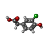  Mass: 186.592 Da / Num. of mol.: 1 / Source method: obtained synthetically / Formula: C8H7ClO3 / Feature type: SUBJECT OF INVESTIGATION Mass: 186.592 Da / Num. of mol.: 1 / Source method: obtained synthetically / Formula: C8H7ClO3 / Feature type: SUBJECT OF INVESTIGATION |
| Has ligand of interest | Y |
| Has protein modification | Y |
-Experimental details
-Experiment
| Experiment | Method: ELECTRON MICROSCOPY |
|---|---|
| EM experiment | Aggregation state: PARTICLE / 3D reconstruction method: single particle reconstruction |
- Sample preparation
| Component | Name: AUX1 protein in complex with CHPAA / Type: COMPLEX / Entity ID: #1 / Source: RECOMBINANT |
|---|---|
| Source (natural) | Organism: |
| Source (recombinant) | Organism: Spodoptera frugiperda (fall armyworm) |
| Buffer solution | pH: 7.4 |
| Specimen | Embedding applied: NO / Shadowing applied: NO / Staining applied: NO / Vitrification applied: YES |
| Vitrification | Cryogen name: ETHANE |
- Electron microscopy imaging
| Experimental equipment |  Model: Titan Krios / Image courtesy: FEI Company |
|---|---|
| Microscopy | Model: TFS KRIOS |
| Electron gun | Electron source:  FIELD EMISSION GUN / Accelerating voltage: 300 kV / Illumination mode: OTHER FIELD EMISSION GUN / Accelerating voltage: 300 kV / Illumination mode: OTHER |
| Electron lens | Mode: BRIGHT FIELD / Nominal defocus max: 2000 nm / Nominal defocus min: 1000 nm |
| Image recording | Electron dose: 50 e/Å2 / Film or detector model: GATAN K3 BIOQUANTUM (6k x 4k) |
- Processing
| EM software | Name: PHENIX / Version: 1.13_2998: / Category: model refinement |
|---|---|
| CTF correction | Type: NONE |
| 3D reconstruction | Resolution: 3.4 Å / Resolution method: FSC 0.143 CUT-OFF / Num. of particles: 239360 / Symmetry type: POINT |
