+ データを開く
- 基本情報
基本情報
| 登録情報 | データベース: PDB / ID: 9lx5 | ||||||
|---|---|---|---|---|---|---|---|
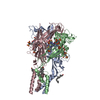
| タイトル | Cryo-EM structure of the P2X1 receptor bound to ATP | ||||||
 要素 要素 | P2X purinoceptor 1 | ||||||
 キーワード キーワード | MEMBRANE PROTEIN / Ion channel | ||||||
| 機能・相同性 |  機能・相同性情報 機能・相同性情報Platelet homeostasis / insemination / positive regulation of calcium ion import across plasma membrane / extracellularly ATP-gated monoatomic cation channel activity / purinergic nucleotide receptor activity / suramin binding / regulation of vascular associated smooth muscle contraction / ligand-gated calcium channel activity / serotonin secretion by platelet / Elevation of cytosolic Ca2+ levels ...Platelet homeostasis / insemination / positive regulation of calcium ion import across plasma membrane / extracellularly ATP-gated monoatomic cation channel activity / purinergic nucleotide receptor activity / suramin binding / regulation of vascular associated smooth muscle contraction / ligand-gated calcium channel activity / serotonin secretion by platelet / Elevation of cytosolic Ca2+ levels / regulation of presynaptic cytosolic calcium ion concentration / ceramide biosynthetic process / response to ATP / regulation of synaptic vesicle exocytosis / neuronal action potential / monoatomic cation channel activity / specific granule membrane / monoatomic ion transport / presynaptic active zone membrane / secretory granule membrane / synaptic transmission, glutamatergic / calcium ion transmembrane transport / platelet activation / regulation of blood pressure / postsynaptic membrane / membrane raft / external side of plasma membrane / apoptotic process / Neutrophil degranulation / protein-containing complex binding / glutamatergic synapse / signal transduction / protein-containing complex / ATP binding / identical protein binding / plasma membrane 類似検索 - 分子機能 | ||||||
| 生物種 |  Homo sapiens (ヒト) Homo sapiens (ヒト) | ||||||
| 手法 | 電子顕微鏡法 / 単粒子再構成法 / クライオ電子顕微鏡法 / 解像度: 3.6 Å | ||||||
 データ登録者 データ登録者 | Qiang, Y. / Chen, K. | ||||||
| 資金援助 |  中国, 1件 中国, 1件
| ||||||
 引用 引用 |  ジャーナル: Acta Pharmacol Sin / 年: 2025 ジャーナル: Acta Pharmacol Sin / 年: 2025タイトル: Structural basis of the multiple ligand binding mechanisms of the P2X1 receptor. 著者: Yu-Ting Qiang / Peng-Peng Wu / Xin Liu / Li Peng / Li-Ke Zhao / Ya-Ting Chen / Zhao-Bing Gao / Qiang Zhao / Kun Chen /  要旨: As important modulators of human purinergic signaling, P2X1 receptors form homotrimers to transport calcium, regulating multiple physiological processes, and are long regarded as promising ...As important modulators of human purinergic signaling, P2X1 receptors form homotrimers to transport calcium, regulating multiple physiological processes, and are long regarded as promising therapeutic targets for male contraception and inflammation. However, the development of drugs that target the P2X1 receptor, such as the antagonist NF449, is greatly hindered by the unclear molecular mechanism of ligand binding modes and receptor activation. Here, we report the structures of the P2X1 receptor in complex with the endogenous agonist ATP or the competitive antagonist NF449. The P2X1 receptor displays distinct conformational features when bound to different types of compounds. Despite coupling to the agonist ATP, the receptor adopts a desensitized conformation that arrests the ions in the transmembrane (TM) domain, aligning with the nature of the high desensitization rates of the P2X1 receptor within the P2X family. Interestingly, the antagonist NF449 not only occupies the orthosteric pocket of ATP but also interacts with the dorsal fin, left flipper, and head domains, suggesting a unique binding mode to perform both orthosteric and allosteric mechanisms of NF449 inhibition. Intriguingly, a novel lipid binding site adjacent to the TM helices and lower body of P2X1, which is critical for receptor activation, is identified. Further functional assay results and structural alignments reveal the high conservation of this lipid binding site in P2X receptors, indicating important modulatory roles upon lipid binding. Taken together, these findings greatly increase our understanding of the ligand binding modes and multiple modulatory mechanisms of the P2X1 receptor and shed light on the further development of P2X1-selective antagonists.Keywords: Structural biology; Ligand binding mode; Ion channel; Purinergic P2X1 receptor. | ||||||
| 履歴 |
|
- 構造の表示
構造の表示
| 構造ビューア | 分子:  Molmil Molmil Jmol/JSmol Jmol/JSmol |
|---|
- ダウンロードとリンク
- ダウンロード
| PDBx/mmCIF形式 |  9lx5.cif.gz 9lx5.cif.gz | 179.3 KB | 表示 |  PDBx/mmCIF形式 PDBx/mmCIF形式 |
|---|---|---|---|---|
| PDB形式 |  pdb9lx5.ent.gz pdb9lx5.ent.gz | 141.1 KB | 表示 |  PDB形式 PDB形式 |
| PDBx/mmJSON形式 |  9lx5.json.gz 9lx5.json.gz | ツリー表示 |  PDBx/mmJSON形式 PDBx/mmJSON形式 | |
| その他 |  その他のダウンロード その他のダウンロード |
-検証レポート
| 文書・要旨 |  9lx5_validation.pdf.gz 9lx5_validation.pdf.gz | 711.6 KB | 表示 |  wwPDB検証レポート wwPDB検証レポート |
|---|---|---|---|---|
| 文書・詳細版 |  9lx5_full_validation.pdf.gz 9lx5_full_validation.pdf.gz | 717.8 KB | 表示 | |
| XML形式データ |  9lx5_validation.xml.gz 9lx5_validation.xml.gz | 21.6 KB | 表示 | |
| CIF形式データ |  9lx5_validation.cif.gz 9lx5_validation.cif.gz | 31.6 KB | 表示 | |
| アーカイブディレクトリ |  https://data.pdbj.org/pub/pdb/validation_reports/lx/9lx5 https://data.pdbj.org/pub/pdb/validation_reports/lx/9lx5 ftp://data.pdbj.org/pub/pdb/validation_reports/lx/9lx5 ftp://data.pdbj.org/pub/pdb/validation_reports/lx/9lx5 | HTTPS FTP |
-関連構造データ
| 関連構造データ |  63466MC  9lxcC M: このデータのモデリングに利用したマップデータ C: 同じ文献を引用 ( |
|---|---|
| 類似構造データ | 類似検索 - 機能・相同性  F&H 検索 F&H 検索 |
- リンク
リンク
- 集合体
集合体
| 登録構造単位 | 
|
|---|---|
| 1 |
|
- 要素
要素
| #1: タンパク質 | 分子量: 36952.723 Da / 分子数: 3 / 由来タイプ: 組換発現 / 由来: (組換発現)  Homo sapiens (ヒト) / 遺伝子: P2RX1, P2X1 Homo sapiens (ヒト) / 遺伝子: P2RX1, P2X1発現宿主:  参照: UniProt: P51575 #2: 化合物 | #3: 化合物 | 研究の焦点であるリガンドがあるか | Y | Has protein modification | Y | |
|---|
-実験情報
-実験
| 実験 | 手法: 電子顕微鏡法 |
|---|---|
| EM実験 | 試料の集合状態: PARTICLE / 3次元再構成法: 単粒子再構成法 |
- 試料調製
試料調製
| 構成要素 | 名称: the ATP-bound P2X1 receptor / タイプ: COMPLEX / Entity ID: #1 / 由来: RECOMBINANT |
|---|---|
| 分子量 | 実験値: NO |
| 由来(天然) | 生物種:  Homo sapiens (ヒト) Homo sapiens (ヒト) |
| 由来(組換発現) | 生物種:  |
| 緩衝液 | pH: 8 |
| 試料 | 濃度: 7 mg/ml / 包埋: NO / シャドウイング: NO / 染色: NO / 凍結: YES |
| 急速凍結 | 凍結剤: ETHANE |
- 電子顕微鏡撮影
| 実験機器 |  モデル: Titan Krios / 画像提供: FEI Company |
|---|---|
| 顕微鏡 | モデル: TFS KRIOS |
| 電子銃 | 電子線源:  FIELD EMISSION GUN / 加速電圧: 300 kV / 照射モード: FLOOD BEAM FIELD EMISSION GUN / 加速電圧: 300 kV / 照射モード: FLOOD BEAM |
| 電子レンズ | モード: BRIGHT FIELD / 最大 デフォーカス(公称値): 1500 nm / 最小 デフォーカス(公称値): 800 nm |
| 撮影 | 電子線照射量: 70 e/Å2 フィルム・検出器のモデル: GATAN K3 BIOQUANTUM (6k x 4k) |
- 解析
解析
| EMソフトウェア | 名称: PHENIX / カテゴリ: モデル精密化 | ||||||||||||||||||||||||
|---|---|---|---|---|---|---|---|---|---|---|---|---|---|---|---|---|---|---|---|---|---|---|---|---|---|
| CTF補正 | タイプ: NONE | ||||||||||||||||||||||||
| 3次元再構成 | 解像度: 3.6 Å / 解像度の算出法: FSC 0.143 CUT-OFF / 粒子像の数: 346074 / 対称性のタイプ: POINT | ||||||||||||||||||||||||
| 原子モデル構築 | プロトコル: AB INITIO MODEL | ||||||||||||||||||||||||
| 精密化 | 最高解像度: 3.6 Å 立体化学のターゲット値: REAL-SPACE (WEIGHTED MAP SUM AT ATOM CENTERS) | ||||||||||||||||||||||||
| 拘束条件 |
|
 ムービー
ムービー コントローラー


 PDBj
PDBj


