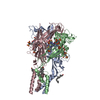
Journal: Acta Pharmacol Sin / Year: 2025 Title: Structural basis of the multiple ligand binding mechanisms of the P2X1 receptor. Authors: Yu-Ting Qiang / Peng-Peng Wu / Xin Liu / Li Peng / Li-Ke Zhao / Ya-Ting Chen / Zhao-Bing Gao / Qiang Zhao / Kun Chen / Abstract: As important modulators of human purinergic signaling, P2X1 receptors form homotrimers to transport calcium, regulating multiple physiological processes, and are long regarded as promising ...As important modulators of human purinergic signaling, P2X1 receptors form homotrimers to transport calcium, regulating multiple physiological processes, and are long regarded as promising therapeutic targets for male contraception and inflammation. However, the development of drugs that target the P2X1 receptor, such as the antagonist NF449, is greatly hindered by the unclear molecular mechanism of ligand binding modes and receptor activation. Here, we report the structures of the P2X1 receptor in complex with the endogenous agonist ATP or the competitive antagonist NF449. The P2X1 receptor displays distinct conformational features when bound to different types of compounds. Despite coupling to the agonist ATP, the receptor adopts a desensitized conformation that arrests the ions in the transmembrane (TM) domain, aligning with the nature of the high desensitization rates of the P2X1 receptor within the P2X family. Interestingly, the antagonist NF449 not only occupies the orthosteric pocket of ATP but also interacts with the dorsal fin, left flipper, and head domains, suggesting a unique binding mode to perform both orthosteric and allosteric mechanisms of NF449 inhibition. Intriguingly, a novel lipid binding site adjacent to the TM helices and lower body of P2X1, which is critical for receptor activation, is identified. Further functional assay results and structural alignments reveal the high conservation of this lipid binding site in P2X receptors, indicating important modulatory roles upon lipid binding. Taken together, these findings greatly increase our understanding of the ligand binding modes and multiple modulatory mechanisms of the P2X1 receptor and shed light on the further development of P2X1-selective antagonists.Keywords: Structural biology; Ligand binding mode; Ion channel; Purinergic P2X1 receptor.
In the structure databanks used in Yorodumi, some data are registered as the other names, "COVID-19 virus" and "2019-nCoV". Here are the details of the virus and the list of structure data.
Jan 31, 2019. EMDB accession codes are about to change! (news from PDBe EMDB page)
EMDB accession codes are about to change! (news from PDBe EMDB page)
The allocation of 4 digits for EMDB accession codes will soon come to an end. Whilst these codes will remain in use, new EMDB accession codes will include an additional digit and will expand incrementally as the available range of codes is exhausted. The current 4-digit format prefixed with “EMD-” (i.e. EMD-XXXX) will advance to a 5-digit format (i.e. EMD-XXXXX), and so on. It is currently estimated that the 4-digit codes will be depleted around Spring 2019, at which point the 5-digit format will come into force.
The EM Navigator/Yorodumi systems omit the EMD- prefix.
Related info.:Q: What is EMD? / ID/Accession-code notation in Yorodumi/EM Navigator
Yorodumi is a browser for structure data from EMDB, PDB, SASBDB, etc.
This page is also the successor to EM Navigator detail page, and also detail information page/front-end page for Omokage search.
The word "yorodu" (or yorozu) is an old Japanese word meaning "ten thousand". "mi" (miru) is to see.
Related info.:EMDB / PDB / SASBDB / Comparison of 3 databanks / Yorodumi Search / Aug 31, 2016. New EM Navigator & Yorodumi / Yorodumi Papers / Jmol/JSmol / Function and homology information / Changes in new EM Navigator and Yorodumi
 Movie
Movie Controller


 Open data
 Basic information
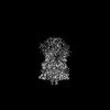
 Map data
 Sample
 Keywords
 Function and homology information
 Homo sapiens (human)
 Authors
 China, 1 items
  Citation
 Structure visualization
 Downloads & links
 emd_63466.png
 http://ftp.pdbj.org/pub/emdb/structures/EMD-63466
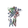
 Z (Sec.)
 Y (Row.)
 X (Col.)


 Sample components
 Spodoptera frugiperda (fall armyworm)

 Processing
 Electron microscopy
 FIELD EMISSION GUN
