 Movie
Movie Controller


+ Open data

- Basic information
| Entry | Database: PDB / ID: 9lsz | |||||||||||||||||||||
|---|---|---|---|---|---|---|---|---|---|---|---|---|---|---|---|---|---|---|---|---|---|---|
| Title | Inward-open Structure of human B0AT1 | |||||||||||||||||||||
 Components Components |
| |||||||||||||||||||||
 Keywords Keywords | TRANSPORT PROTEIN / amino acid transporter / SLC6 | |||||||||||||||||||||
| Function / homology |  Function and homology information Function and homology informationDefective transport of neurotransmitters by SLC6A19 causes Hartnup disorder (HND) / Defective transport of amino acids by SLC6A19 causes Hartnup disorder (HND) / neutral amino acid transport / neutral L-amino acid transmembrane transporter activity / symporter activity / Amino acid transport across the plasma membrane / amino acid transmembrane transporter activity / SLC-mediated transport of neurotransmitters / neurotransmitter transport / amino acid transport ...Defective transport of neurotransmitters by SLC6A19 causes Hartnup disorder (HND) / Defective transport of amino acids by SLC6A19 causes Hartnup disorder (HND) / neutral amino acid transport / neutral L-amino acid transmembrane transporter activity / symporter activity / Amino acid transport across the plasma membrane / amino acid transmembrane transporter activity / SLC-mediated transport of neurotransmitters / neurotransmitter transport / amino acid transport / amino acid import across plasma membrane / viral life cycle / response to nutrient / sodium ion transmembrane transport / brush border membrane / apical plasma membrane / extracellular exosome / plasma membrane Similarity search - Function | |||||||||||||||||||||
| Biological species |  Homo sapiens (human) Homo sapiens (human) | |||||||||||||||||||||
| Method | ELECTRON MICROSCOPY / single particle reconstruction / cryo EM / Resolution: 3.11 Å | |||||||||||||||||||||
 Authors Authors | Imazu, T. / Miyaguchi, I. / Hiraizumi, M. | |||||||||||||||||||||
| Funding support | 1items
| |||||||||||||||||||||
 Citation Citation | Journal: To Be Published Title: Structure-guided development of a potent human B0AT1 inhibitor effective in a mouse model of phenylketonuria Authors: Imazu, T. / Akashi, T. / Hiraizumi, M. / Inui, Y. / Sasaki, W. / Takahashi, T. / Todoroki, H. / Kumanomidou, T. / Hisano, H. / Asada, H. / Kusakizako, T. / Nishizawa, T. / Iwata, S. / Nureki, O. / Miyaguchi, I. | |||||||||||||||||||||
| History |
|
- Structure visualization
| Structure viewer | Molecule: MolmilJmol/JSmol |
|---|
- Downloads & links
-Download
| PDBx/mmCIF format | 9lsz.cif.gz | 183.9 KB | Display | PDBx/mmCIF format |
|---|---|---|---|---|
| PDB format | pdb9lsz.ent.gz | 136.9 KB | Display | PDB format |
| PDBx/mmJSON format | 9lsz.json.gz | Tree view | PDBx/mmJSON format | |
| Others |  Other downloads Other downloads |
-Validation report
| Arichive directory | https://data.pdbj.org/pub/pdb/validation_reports/ls/9lszftp://data.pdbj.org/pub/pdb/validation_reports/ls/9lsz | HTTPS FTP |
|---|
-Related structure data
| Related structure data |  63360MC 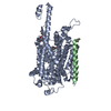 9kxtC  9kxuC  9kxvC  9kxwC  9kxxC  9kxyC C: citing same article ( M: map data used to model this data |
|---|---|
| Similar structure data |


-Links
 PDBj
PDBj


- Assembly
| Deposited unit | 
|
|---|---|
| 1 |
|
-Components
| #1: Protein | Mass: 68874.625 Da / Num. of mol.: 1 Source method: isolated from a genetically manipulated source Source: (gene. exp.) Homo sapiens (human) / Gene: SLC6A19, B0AT1 / Production host: Homo sapiens (human) / References: UniProt: Q695T7 |
|---|---|
| #2: Antibody | Mass: 24845.598 Da / Num. of mol.: 1 / Source method: isolated from a natural source / Source: (natural) |
| #3: Antibody | Mass: 36524.039 Da / Num. of mol.: 1 / Source method: isolated from a natural source / Source: (natural) |
| #4: Polysaccharide | 2-acetamido-2-deoxy-beta-D-glucopyranose-(1-4)-2-acetamido-2-deoxy-beta-D-glucopyranose Source method: isolated from a genetically manipulated source |
| Has ligand of interest | N |
| Has protein modification | Y |
-Experimental details
-Experiment
| Experiment | Method: ELECTRON MICROSCOPY |
|---|---|
| EM experiment | Aggregation state: PARTICLE / 3D reconstruction method: single particle reconstruction |
- Sample preparation
| Component | Name: Binary complex of B0AT1 and antibody / Type: COMPLEX / Entity ID: #1-#3 / Source: RECOMBINANT |
|---|---|
| Molecular weight | Value: 0.12 MDa / Experimental value: NO |
| Source (natural) | Organism: Homo sapiens (human) |
| Source (recombinant) | Organism: Homo sapiens (human) |
| Buffer solution | pH: 7.5 |
| Specimen | Embedding applied: NO / Shadowing applied: NO / Staining applied: NO / Vitrification applied: YES |
| Vitrification | Cryogen name: ETHANE |
- Electron microscopy imaging
| Experimental equipment |  Model: Titan Krios / Image courtesy: FEI Company |
|---|---|
| Microscopy | Model: TFS KRIOS |
| Electron gun | Electron source:  FIELD EMISSION GUN / Accelerating voltage: 300 kV / Illumination mode: FLOOD BEAM FIELD EMISSION GUN / Accelerating voltage: 300 kV / Illumination mode: FLOOD BEAM |
| Electron lens | Mode: BRIGHT FIELD / Nominal defocus max: 1600 nm / Nominal defocus min: 800 nm |
| Image recording | Electron dose: 7.6 e/Å2 / Film or detector model: GATAN K3 (6k x 4k) |
- Processing
| EM software | Name: REFMAC / Version: 5.8.0267 / Category: model refinement | ||||||||||||||||||||||||||||||||||||||||||||||||||||||||||||||||||||||||||||||||||||||||||||||||||||||||||
|---|---|---|---|---|---|---|---|---|---|---|---|---|---|---|---|---|---|---|---|---|---|---|---|---|---|---|---|---|---|---|---|---|---|---|---|---|---|---|---|---|---|---|---|---|---|---|---|---|---|---|---|---|---|---|---|---|---|---|---|---|---|---|---|---|---|---|---|---|---|---|---|---|---|---|---|---|---|---|---|---|---|---|---|---|---|---|---|---|---|---|---|---|---|---|---|---|---|---|---|---|---|---|---|---|---|---|---|
| CTF correction | Type: PHASE FLIPPING AND AMPLITUDE CORRECTION | ||||||||||||||||||||||||||||||||||||||||||||||||||||||||||||||||||||||||||||||||||||||||||||||||||||||||||
| 3D reconstruction | Resolution: 3.11 Å / Resolution method: FSC 0.143 CUT-OFF / Num. of particles: 407749 / Symmetry type: POINT | ||||||||||||||||||||||||||||||||||||||||||||||||||||||||||||||||||||||||||||||||||||||||||||||||||||||||||
| Refinement | Resolution: 3.11→3.11 Å / Cor.coef. Fo:Fc: 0.905 / SU B: 9.133 / SU ML: 0.171 / ESU R: 0.165 Stereochemistry target values: MAXIMUM LIKELIHOOD WITH PHASES Details: HYDROGENS HAVE BEEN ADDED IN THE RIDING POSITIONS
| ||||||||||||||||||||||||||||||||||||||||||||||||||||||||||||||||||||||||||||||||||||||||||||||||||||||||||
| Solvent computation | Solvent model: PARAMETERS FOR MASK CACLULATION | ||||||||||||||||||||||||||||||||||||||||||||||||||||||||||||||||||||||||||||||||||||||||||||||||||||||||||
| Displacement parameters | Biso mean: 117.002 Å2 | ||||||||||||||||||||||||||||||||||||||||||||||||||||||||||||||||||||||||||||||||||||||||||||||||||||||||||
| Refinement step | Cycle: 1 / Total: 6458 | ||||||||||||||||||||||||||||||||||||||||||||||||||||||||||||||||||||||||||||||||||||||||||||||||||||||||||
| Refine LS restraints |
|