 ムービー
ムービー コントローラー


+ データを開く

- 基本情報
基本情報
| 登録情報 | データベース: PDB / ID: 9kzl | ||||||
|---|---|---|---|---|---|---|---|
| タイトル | Crystal structure of the functional unit (HtH1-h) of hemocyanin from Haliotis discus hannai,Containing a met site | ||||||
 要素 要素 | (Hemocyanin type ...) x 2 | ||||||
 キーワード キーワード | METAL BINDING PROTEIN / Orbicular / copper coordination / Met state | ||||||
| 機能・相同性 |  機能・相同性情報 機能・相同性情報 | ||||||
| 生物種 |  Haliotis discus hannai (エゾアワビ) Haliotis discus hannai (エゾアワビ) | ||||||
| 手法 |  X線回折 / シンクロトロン / 分子置換 / 解像度: 2 Å X線回折 / シンクロトロン / 分子置換 / 解像度: 2 Å | ||||||
 データ登録者 データ登録者 | Zhao, G.H. / Yang, H.C. | ||||||
| 資金援助 |  中国, 1件 中国, 1件
| ||||||
 引用 引用 | ジャーナル: To Be Published タイトル: Crystal structure of the functional unit (HtH1-h) of hemocyanin from Haliotis discus hannai,Containing a met state 著者: Zhao, G.H. / Yang, H.C. | ||||||
| 履歴 |
|
- 構造の表示
構造の表示
| 構造ビューア | 分子: MolmilJmol/JSmol |
|---|
- ダウンロードとリンク
-ダウンロード
| PDBx/mmCIF形式 | 9kzl.cif.gz | 411.8 KB | 表示 | PDBx/mmCIF形式 |
|---|---|---|---|---|
| PDB形式 | pdb9kzl.ent.gz | 267.1 KB | 表示 | PDB形式 |
| PDBx/mmJSON形式 | 9kzl.json.gz | ツリー表示 | PDBx/mmJSON形式 | |
| その他 |  その他のダウンロード その他のダウンロード |
-検証レポート
| 文書・要旨 | 9kzl_validation.pdf.gz | 7.6 MB | 表示 | wwPDB検証レポート |
|---|---|---|---|---|
| 文書・詳細版 | 9kzl_full_validation.pdf.gz | 7.6 MB | 表示 | |
| XML形式データ | 9kzl_validation.xml.gz | 75.9 KB | 表示 | |
| CIF形式データ | 9kzl_validation.cif.gz | 104 KB | 表示 | |
| アーカイブディレクトリ | https://data.pdbj.org/pub/pdb/validation_reports/kz/9kzlftp://data.pdbj.org/pub/pdb/validation_reports/kz/9kzl | HTTPS FTP |
-関連構造データ
| 関連構造データ | 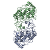 3qjoS S: 精密化の開始モデル |
|---|---|
| 類似構造データ |
-リンク
 PDBj
PDBj


- 集合体
集合体
| 登録構造単位 | 
| ||||||||||||
|---|---|---|---|---|---|---|---|---|---|---|---|---|---|
| 1 | 
| ||||||||||||
| 2 | 
| ||||||||||||
| 単位格子 |
|
-要素
-Hemocyanin type ... , 2種, 3分子 ABC
| #1: タンパク質 | 分子量: 56653.176 Da / 分子数: 1 / 由来タイプ: 天然 / 由来: (天然) Haliotis discus hannai (エゾアワビ) / 参照: UniProt: A0A7R6RYX0 |
|---|---|
| #2: タンパク質 | 分子量: 56639.148 Da / 分子数: 2 / 由来タイプ: 天然 / 由来: (天然) Haliotis discus hannai (エゾアワビ) / 参照: UniProt: A0A7R6RYX0 |
-糖 , 1種, 3分子 
| #3: 糖 |  タイプ: D-saccharide, alpha linking / 分子量: 180.156 Da / 分子数: 3 / 由来タイプ: 合成 / 式: C6H12O6 / タイプ: SUBJECT OF INVESTIGATION タイプ: D-saccharide, alpha linking / 分子量: 180.156 Da / 分子数: 3 / 由来タイプ: 合成 / 式: C6H12O6 / タイプ: SUBJECT OF INVESTIGATION |
|---|
-非ポリマー , 5種, 1116分子 


| #4: 化合物 |  分子量: 46.068 Da / 分子数: 3 / 由来タイプ: 合成 / 式: C2H6O / タイプ: SUBJECT OF INVESTIGATION 分子量: 46.068 Da / 分子数: 3 / 由来タイプ: 合成 / 式: C2H6O / タイプ: SUBJECT OF INVESTIGATION#5: 化合物 | ChemComp-CU /  分子量: 63.546 Da / 分子数: 6 / 由来タイプ: 合成 / 式: Cu / タイプ: SUBJECT OF INVESTIGATION 分子量: 63.546 Da / 分子数: 6 / 由来タイプ: 合成 / 式: Cu / タイプ: SUBJECT OF INVESTIGATION#6: 化合物 |  分子量: 17.007 Da / 分子数: 2 / 由来タイプ: 合成 / 式: HO / タイプ: SUBJECT OF INVESTIGATION 分子量: 17.007 Da / 分子数: 2 / 由来タイプ: 合成 / 式: HO / タイプ: SUBJECT OF INVESTIGATION#7: 化合物 |  分子量: 15.999 Da / 分子数: 2 / 由来タイプ: 合成 / 式: O / タイプ: SUBJECT OF INVESTIGATION 分子量: 15.999 Da / 分子数: 2 / 由来タイプ: 合成 / 式: O / タイプ: SUBJECT OF INVESTIGATION#8: 水 | ChemComp-HOH / | 分子量: 18.015 Da / 分子数: 1103 / 由来タイプ: 天然 / 式: H2O |
|---|
-詳細
| 研究の焦点であるリガンドがあるか | Y |
|---|---|
| Has protein modification | Y |
-実験情報
-実験
| 実験 | 手法: X線回折 / 使用した結晶の数: 1 |
|---|
- 試料調製
試料調製
| 結晶 | マシュー密度: 2.83 Å3/Da / 溶媒含有率: 56.52 % |
|---|---|
| 結晶化 | 温度: 293.15 K / 手法: 蒸気拡散法, シッティングドロップ法 / pH: 4.2 / 詳細: PEG 1000,Ethanol,Phosphate |
-データ収集
| 回折 | 平均測定温度: 283 K / Serial crystal experiment: N |
|---|---|
| 放射光源 | 由来: シンクロトロン / サイト: SSRF / ビームライン: BL18U1 / 波長: 0.987 Å |
| 検出器 | タイプ: DECTRIS PILATUS3 6M / 検出器: PIXEL / 日付: 2023年11月17日 |
| 放射 | モノクロメーター: Si111 / プロトコル: SINGLE WAVELENGTH / 単色(M)・ラウエ(L): M / 散乱光タイプ: x-ray |
| 放射波長 | 波長: 0.987 Å / 相対比: 1 |
| 反射 | 解像度: 1.999→28.6 Å / Num. obs: 126200 / % possible obs: 98.86 % / 冗長度: 3.5 % / Biso Wilson estimate: 26.01 Å2 / CC1/2: 0.98 / Net I/σ(I): 4.3 |
| 反射 シェル | 解像度: 2→2.02 Å / Mean I/σ(I) obs: 14.9 / Num. unique obs: 3720 / CC1/2: 0.988 / % possible all: 92.58 |
- 解析
解析
| ソフトウェア |
| |||||||||||||||||||||||||||||||||||||||||||||||||||||||||||||||||||||||||||||||||||||||||||||||||||||||||||||||||||||||||||||||||||||||||||||||||||||||||||||||||||||||||||||||||||||||||||||||||||||||||||||||||||||||||
|---|---|---|---|---|---|---|---|---|---|---|---|---|---|---|---|---|---|---|---|---|---|---|---|---|---|---|---|---|---|---|---|---|---|---|---|---|---|---|---|---|---|---|---|---|---|---|---|---|---|---|---|---|---|---|---|---|---|---|---|---|---|---|---|---|---|---|---|---|---|---|---|---|---|---|---|---|---|---|---|---|---|---|---|---|---|---|---|---|---|---|---|---|---|---|---|---|---|---|---|---|---|---|---|---|---|---|---|---|---|---|---|---|---|---|---|---|---|---|---|---|---|---|---|---|---|---|---|---|---|---|---|---|---|---|---|---|---|---|---|---|---|---|---|---|---|---|---|---|---|---|---|---|---|---|---|---|---|---|---|---|---|---|---|---|---|---|---|---|---|---|---|---|---|---|---|---|---|---|---|---|---|---|---|---|---|---|---|---|---|---|---|---|---|---|---|---|---|---|---|---|---|---|---|---|---|---|---|---|---|---|---|---|---|---|---|---|---|---|
| 精密化 | 構造決定の手法: 分子置換 開始モデル: 3QJO 解像度: 2→28.6 Å / SU ML: 0.1961 / 交差検証法: FREE R-VALUE / σ(F): 1.34 / 位相誤差: 21.2398 立体化学のターゲット値: GeoStd + Monomer Library + CDL v1.2
| |||||||||||||||||||||||||||||||||||||||||||||||||||||||||||||||||||||||||||||||||||||||||||||||||||||||||||||||||||||||||||||||||||||||||||||||||||||||||||||||||||||||||||||||||||||||||||||||||||||||||||||||||||||||||
| 溶媒の処理 | 減衰半径: 0.9 Å / VDWプローブ半径: 1.1 Å / 溶媒モデル: FLAT BULK SOLVENT MODEL | |||||||||||||||||||||||||||||||||||||||||||||||||||||||||||||||||||||||||||||||||||||||||||||||||||||||||||||||||||||||||||||||||||||||||||||||||||||||||||||||||||||||||||||||||||||||||||||||||||||||||||||||||||||||||
| 原子変位パラメータ | Biso mean: 29.14 Å2 | |||||||||||||||||||||||||||||||||||||||||||||||||||||||||||||||||||||||||||||||||||||||||||||||||||||||||||||||||||||||||||||||||||||||||||||||||||||||||||||||||||||||||||||||||||||||||||||||||||||||||||||||||||||||||
| 精密化ステップ | サイクル: LAST / 解像度: 2→28.6 Å
| |||||||||||||||||||||||||||||||||||||||||||||||||||||||||||||||||||||||||||||||||||||||||||||||||||||||||||||||||||||||||||||||||||||||||||||||||||||||||||||||||||||||||||||||||||||||||||||||||||||||||||||||||||||||||
| 拘束条件 |
| |||||||||||||||||||||||||||||||||||||||||||||||||||||||||||||||||||||||||||||||||||||||||||||||||||||||||||||||||||||||||||||||||||||||||||||||||||||||||||||||||||||||||||||||||||||||||||||||||||||||||||||||||||||||||
| LS精密化 シェル |
|
