 Movie
Movie Controller

[English] 日本語
 Yorodumi
- PDB-9kn8: Crystal structure of Horse spleen L-ferritin mutant (E53F/E56F/E5... -
+ Open data

- Basic information
| Entry | Database: PDB / ID: 9kn8 | ||||||
|---|---|---|---|---|---|---|---|
| Title | Crystal structure of Horse spleen L-ferritin mutant (E53F/E56F/E57F/R59F/E60F/E63F) with Coumarin 153 | ||||||
 Components Components | Ferritin light chain | ||||||
 Keywords Keywords | METAL BINDING PROTEIN / 24mer cage / Phenyl Alanine mutant / Coumarin 153 | ||||||
| Function / homology |  Function and homology information Function and homology informationferritin complex / autolysosome / ferric iron binding / autophagosome / iron ion transport / ferrous iron binding / cytoplasmic vesicle / intracellular iron ion homeostasis / iron ion binding / cytoplasm Similarity search - Function | ||||||
| Biological species |  | ||||||
| Method |  X-RAY DIFFRACTION / MOLECULAR REPLACEMENT / Resolution: 1.64 Å X-RAY DIFFRACTION / MOLECULAR REPLACEMENT / Resolution: 1.64 Å | ||||||
 Authors Authors | Suzuki, T. / Hishikawa, Y. / Maity, B. / Abe, S. / Ueno, T. | ||||||
| Funding support |  Japan, 1items Japan, 1items
| ||||||
 Citation Citation | Journal: Adv Sci / Year: 2025 Title: Design of Aromatic Interaction Networks in a Protein Cage Modulated by Fluorescent Ligand Binding. Authors: Hishikawa, Y. / Suzuki, T. / Maity, B. / Noya, H. / Yoshizawa, M. / Asanuma, A. / Katagiri, Y. / Abe, S. / Nagatoishi, S. / Tsumoto, K. / Ueno, T. | ||||||
| History |
|
- Structure visualization
| Structure viewer | Molecule: MolmilJmol/JSmol |
|---|
- Downloads & links
-Download
| PDBx/mmCIF format | 9kn8.cif.gz | 57.8 KB | Display | PDBx/mmCIF format |
|---|---|---|---|---|
| PDB format | pdb9kn8.ent.gz | Display | PDB format | |
| PDBx/mmJSON format | 9kn8.json.gz | Tree view | PDBx/mmJSON format | |
| Others |  Other downloads Other downloads |
-Validation report
| Summary document | 9kn8_validation.pdf.gz | 786.5 KB | Display | wwPDB validaton report |
|---|---|---|---|---|
| Full document | 9kn8_full_validation.pdf.gz | 786.7 KB | Display | |
| Data in XML | 9kn8_validation.xml.gz | 12.7 KB | Display | |
| Data in CIF | 9kn8_validation.cif.gz | 17.5 KB | Display | |
| Arichive directory | https://data.pdbj.org/pub/pdb/validation_reports/kn/9kn8ftp://data.pdbj.org/pub/pdb/validation_reports/kn/9kn8 | HTTPS FTP |
-Related structure data
| Related structure data |  9kkpC  9kkrC  9kleC  9kn7C  9kp2C  9kp5C  9kp7C  9kpaC  9krsC 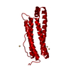 1datS S: Starting model for refinement C: citing same article ( |
|---|---|
| Similar structure data |
-Links
 PDBj
PDBj


- Assembly
| Deposited unit | 
| ||||||||||||||||||
|---|---|---|---|---|---|---|---|---|---|---|---|---|---|---|---|---|---|---|---|
| 1 | x 24
| ||||||||||||||||||
| Unit cell |
| ||||||||||||||||||
| Components on special symmetry positions |
|

-Components
-Protein , 1 types, 1 molecules A
| #1: Protein | Mass: 19952.709 Da / Num. of mol.: 1 / Mutation: E53F/E56F/E57F/R59F/E60F/E63F Source method: isolated from a genetically manipulated source Source: (gene. exp.)  |
|---|
-Non-polymers , 6 types, 208 molecules 


| #2: Chemical | ChemComp-CD /  Mass: 112.411 Da / Num. of mol.: 5 / Source method: obtained synthetically / Formula: Cd Mass: 112.411 Da / Num. of mol.: 5 / Source method: obtained synthetically / Formula: Cd#3: Chemical | ChemComp-SO4 / |  Mass: 96.063 Da / Num. of mol.: 1 / Source method: obtained synthetically / Formula: SO4 Mass: 96.063 Da / Num. of mol.: 1 / Source method: obtained synthetically / Formula: SO4#4: Chemical | ChemComp-A1L54 / | Mass: 309.283 Da / Num. of mol.: 1 / Source method: obtained synthetically / Formula: C16H14F3NO2 / Feature type: SUBJECT OF INVESTIGATION #5: Chemical |  Mass: 62.068 Da / Num. of mol.: 3 / Source method: obtained synthetically / Formula: C2H6O2 Mass: 62.068 Da / Num. of mol.: 3 / Source method: obtained synthetically / Formula: C2H6O2#6: Chemical | ChemComp-CL / |  Mass: 35.453 Da / Num. of mol.: 1 / Source method: obtained synthetically / Formula: Cl Mass: 35.453 Da / Num. of mol.: 1 / Source method: obtained synthetically / Formula: Cl#7: Water | ChemComp-HOH / | Mass: 18.015 Da / Num. of mol.: 197 / Source method: isolated from a natural source / Formula: H2O |
|---|
-Details
| Has ligand of interest | Y |
|---|---|
| Has protein modification | N |
-Experimental details
-Experiment
| Experiment | Method: X-RAY DIFFRACTION / Number of used crystals: 1 |
|---|
- Sample preparation
| Crystal | Density Matthews: 3.1 Å3/Da / Density % sol: 60.37 % |
|---|---|
| Crystal grow | Temperature: 293 K / Method: vapor diffusion, hanging drop / pH: 8 / Details: Ammonium sulfate, Cadmium sulfate / Temp details: Incubation |
-Data collection
| Diffraction | Mean temperature: 93 K / Ambient temp details: Liquid N2 vapour / Serial crystal experiment: N |
|---|---|
| Diffraction source | Source: ROTATING ANODE / Type: RIGAKU FR-E DW / Wavelength: 1.54184 Å |
| Detector | Type: RIGAKU HyPix-6000HE / Detector: PIXEL / Date: Mar 8, 2021 |
| Radiation | Protocol: SINGLE WAVELENGTH / Monochromatic (M) / Laue (L): M / Scattering type: x-ray |
| Radiation wavelength | Wavelength: 1.54184 Å / Relative weight: 1 |
| Reflection | Resolution: 1.64→18.21 Å / Num. obs: 31536 / % possible obs: 99.5 % / Redundancy: 8.4 % / CC1/2: 0.999 / Rmerge(I) obs: 0.071 / Rpim(I) all: 0.024 / Rrim(I) all: 0.075 / Χ2: 0.98 / Net I/σ(I): 20.9 |
| Reflection shell | Resolution: 1.64→1.67 Å / Redundancy: 5.2 % / Rmerge(I) obs: 0.815 / Mean I/σ(I) obs: 2.1 / Num. unique obs: 1587 / CC1/2: 0.819 / Rpim(I) all: 0.378 / Rrim(I) all: 0.903 / Χ2: 1.15 / % possible all: 99.4 |
- Processing
| Software |
| ||||||||||||||||||||||||||||||||||||||||||||||||||||||||||||||||||||||||||||||||||||
|---|---|---|---|---|---|---|---|---|---|---|---|---|---|---|---|---|---|---|---|---|---|---|---|---|---|---|---|---|---|---|---|---|---|---|---|---|---|---|---|---|---|---|---|---|---|---|---|---|---|---|---|---|---|---|---|---|---|---|---|---|---|---|---|---|---|---|---|---|---|---|---|---|---|---|---|---|---|---|---|---|---|---|---|---|---|
| Refinement | Method to determine structure: MOLECULAR REPLACEMENT Starting model: 1DAT Resolution: 1.64→18.21 Å / SU ML: 0.14 / Cross valid method: FREE R-VALUE / σ(F): 1.33 / Phase error: 21.77 / Stereochemistry target values: ML
| ||||||||||||||||||||||||||||||||||||||||||||||||||||||||||||||||||||||||||||||||||||
| Solvent computation | Shrinkage radii: 0.9 Å / VDW probe radii: 1.1 Å / Solvent model: FLAT BULK SOLVENT MODEL | ||||||||||||||||||||||||||||||||||||||||||||||||||||||||||||||||||||||||||||||||||||
| Refinement step | Cycle: LAST / Resolution: 1.64→18.21 Å
| ||||||||||||||||||||||||||||||||||||||||||||||||||||||||||||||||||||||||||||||||||||
| Refine LS restraints |
| ||||||||||||||||||||||||||||||||||||||||||||||||||||||||||||||||||||||||||||||||||||
| LS refinement shell |
|
