 Movie
Movie Controller

[English] 日本語
 Yorodumi
- PDB-9km0: Cryo-EM structure of a tri-heme cytochrome-associated RC-LH1 comp... -
+ Open data

- Basic information
| Entry | Database: PDB / ID: 9km0 | ||||||
|---|---|---|---|---|---|---|---|
| Title | Cryo-EM structure of a tri-heme cytochrome-associated RC-LH1 complex from a marine photoheterotrophic bacterium, purified with EDTA-2Na-containing solutions | ||||||
 Components Components |
| ||||||
 Keywords Keywords | PHOTOSYNTHESIS / Reaction center / energy transfer | ||||||
| Function / homology |  Function and homology information Function and homology informationorganelle inner membrane / plasma membrane-derived chromatophore membrane / plasma membrane light-harvesting complex / bacteriochlorophyll binding / photosynthetic electron transport in photosystem II / photosynthesis, light reaction / electron transfer activity / iron ion binding / heme binding / metal ion binding / plasma membrane Similarity search - Function | ||||||
| Biological species |  Dinoroseobacter shibae DFL 12 = DSM 16493 (bacteria) Dinoroseobacter shibae DFL 12 = DSM 16493 (bacteria) | ||||||
| Method | ELECTRON MICROSCOPY / single particle reconstruction / cryo EM / Resolution: 2.78 Å | ||||||
 Authors Authors | Chen, J.H. | ||||||
| Funding support |  China, 1items China, 1items
| ||||||
 Citation Citation | Journal: Adv Sci (Weinh) / Year: 2025 Title: Cryo-EM Analysis of a Tri-Heme Cytochrome-Associated RC-LH1 Complex from the Marine Photoheterotrophic Bacterium Dinoroseobacter Shibae. Authors: Weiwei Wang / Yanting Liu / Jiayi Gu / Shaoya An / Cheng Ma / Haichun Gao / Nianzhi Jiao / Jian-Ren Shen / John Thomas Beatty / Michal Koblížek / Xing Zhang / Qiang Zheng / Jing-Hua Chen /   Abstract: The reaction center-light harvesting 1 (RC-LH1) complex converts solar energy into electrical energy, driving the initiation of photosynthesis. The authors present a cryo-electron microscopy ...The reaction center-light harvesting 1 (RC-LH1) complex converts solar energy into electrical energy, driving the initiation of photosynthesis. The authors present a cryo-electron microscopy structure of the RC-LH1 isolated from a marine photoheterotrophic bacterium Dinoroseobacter shibae. The RC comprises four subunits, including a three-heme cytochrome (Cyt) c protein, and is surrounded by a closed LH ring composed of 17 pairs of antenna subunits. Notably, a novel subunit with an N-terminal "helix-turn-helix" motif embedded in the gap between the RC and the LH ring is identified. The purified RC-LH1 complex exhibits high stability in solutions containing Mg or Ca. The periplasmic Cyt c is predicted to bind at the junction between the Cyt subunit and the membrane plane, enabling electron transfer from Cyt c to the proximal heme of the tri-heme Cyt, and subsequently to the special pair of bacteriochlorophylls. These findings provide structural insights into the efficient energy and electron transfer processes within a distinct type of RC-LH1, and shed light on evolutionary adaptations of photosynthesis. | ||||||
| History |
|
- Structure visualization
| Structure viewer | Molecule: MolmilJmol/JSmol |
|---|
- Downloads & links
-Download
| PDBx/mmCIF format | 9km0.cif.gz | 638.6 KB | Display | PDBx/mmCIF format |
|---|---|---|---|---|
| PDB format | pdb9km0.ent.gz | Display | PDB format | |
| PDBx/mmJSON format | 9km0.json.gz | Tree view | PDBx/mmJSON format | |
| Others |  Other downloads Other downloads |
-Validation report
| Arichive directory | https://data.pdbj.org/pub/pdb/validation_reports/km/9km0ftp://data.pdbj.org/pub/pdb/validation_reports/km/9km0 | HTTPS FTP |
|---|
-Related structure data
| Related structure data | 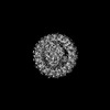 62419MC 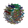 8yy9C 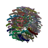 8yz2C M: map data used to model this data C: citing same article ( |
|---|---|
| Similar structure data |
-Links
 PDBj
PDBj


- Assembly
| Deposited unit | 
|
|---|---|
| 1 |
|
-Components
-Antenna pigment protein ... , 2 types, 34 molecules PVSTQR1NKJIGFEDBAvtsrqp2nkjigf...
| #1: Protein | Mass: 6195.409 Da / Num. of mol.: 17 / Source method: isolated from a natural source Source: (natural) Dinoroseobacter shibae DFL 12 = DSM 16493 (bacteria)References: UniProt: A8LQ15 #3: Protein/peptide | Mass: 5578.334 Da / Num. of mol.: 17 / Source method: isolated from a natural source Source: (natural) Dinoroseobacter shibae DFL 12 = DSM 16493 (bacteria)References: UniProt: A8LQ14 |
|---|
-Reaction center protein ... , 4 types, 4 molecules OMLH
| #2: Protein | Mass: 25206.586 Da / Num. of mol.: 1 / Source method: isolated from a natural source Source: (natural) Dinoroseobacter shibae DFL 12 = DSM 16493 (bacteria)References: UniProt: A8LIU2 |
|---|---|
| #4: Protein | Mass: 37583.723 Da / Num. of mol.: 1 / Source method: isolated from a natural source Source: (natural) Dinoroseobacter shibae DFL 12 = DSM 16493 (bacteria)References: UniProt: A8LQ17 |
| #5: Protein | Mass: 31197.178 Da / Num. of mol.: 1 / Source method: isolated from a natural source Source: (natural) Dinoroseobacter shibae DFL 12 = DSM 16493 (bacteria)References: UniProt: A8LQ16 |
| #6: Protein | Mass: 28739.449 Da / Num. of mol.: 1 / Source method: isolated from a natural source Source: (natural) Dinoroseobacter shibae DFL 12 = DSM 16493 (bacteria)References: UniProt: A8LQ33 |
-Protein / Sugars , 2 types, 6 molecules C
| #11: Sugar | ChemComp-LMT /  Type: D-saccharide / Mass: 510.615 Da / Num. of mol.: 5 / Source method: obtained synthetically / Formula: C24H46O11 / Comment: detergent*YM Type: D-saccharide / Mass: 510.615 Da / Num. of mol.: 5 / Source method: obtained synthetically / Formula: C24H46O11 / Comment: detergent*YM#7: Protein | | Mass: 39977.367 Da / Num. of mol.: 1 / Source method: isolated from a natural source Source: (natural) Dinoroseobacter shibae DFL 12 = DSM 16493 (bacteria)References: UniProt: A8LQ18 |
|---|
-Non-polymers , 8 types, 93 molecules 


| #8: Chemical | ChemComp-BCL /  Mass: 911.504 Da / Num. of mol.: 38 / Source method: obtained synthetically / Formula: C55H74MgN4O6 Mass: 911.504 Da / Num. of mol.: 38 / Source method: obtained synthetically / Formula: C55H74MgN4O6#9: Chemical | ChemComp-A1EFU / ( Mass: 582.898 Da / Num. of mol.: 35 / Source method: obtained synthetically / Formula: C41H58O2 / Feature type: SUBJECT OF INVESTIGATION #10: Chemical | ChemComp-MW9 / (  Mass: 777.060 Da / Num. of mol.: 10 / Source method: obtained synthetically / Formula: C42H81O10P Mass: 777.060 Da / Num. of mol.: 10 / Source method: obtained synthetically / Formula: C42H81O10P#12: Chemical | ChemComp-FE / |  Mass: 55.845 Da / Num. of mol.: 1 / Source method: obtained synthetically / Formula: Fe Mass: 55.845 Da / Num. of mol.: 1 / Source method: obtained synthetically / Formula: Fe#13: Chemical |  Mass: 863.343 Da / Num. of mol.: 2 / Source method: obtained synthetically / Formula: C59H90O4 / Feature type: SUBJECT OF INVESTIGATION Mass: 863.343 Da / Num. of mol.: 2 / Source method: obtained synthetically / Formula: C59H90O4 / Feature type: SUBJECT OF INVESTIGATION#14: Chemical |  Mass: 889.215 Da / Num. of mol.: 2 / Source method: obtained synthetically / Formula: C55H76N4O6 Mass: 889.215 Da / Num. of mol.: 2 / Source method: obtained synthetically / Formula: C55H76N4O6#15: Chemical |  Mass: 1464.043 Da / Num. of mol.: 2 / Source method: obtained synthetically / Formula: C81H156O17P2 / Comment: phospholipid*YM Mass: 1464.043 Da / Num. of mol.: 2 / Source method: obtained synthetically / Formula: C81H156O17P2 / Comment: phospholipid*YM#16: Chemical |  Mass: 618.503 Da / Num. of mol.: 3 / Source method: obtained synthetically / Formula: C34H34FeN4O4 / Feature type: SUBJECT OF INVESTIGATION Mass: 618.503 Da / Num. of mol.: 3 / Source method: obtained synthetically / Formula: C34H34FeN4O4 / Feature type: SUBJECT OF INVESTIGATION |
|---|
-Details
| Has ligand of interest | Y |
|---|---|
| Has protein modification | N |
-Experimental details
-Experiment
| Experiment | Method: ELECTRON MICROSCOPY |
|---|---|
| EM experiment | Aggregation state: PARTICLE / 3D reconstruction method: single particle reconstruction |
- Sample preparation
| Component | Name: tri-heme cytochrome-associated RC-LH1 complex / Type: COMPLEX / Entity ID: #1-#7 / Source: NATURAL |
|---|---|
| Source (natural) | Organism: Dinoroseobacter shibae DFL 12 = DSM 16493 (bacteria) |
| Buffer solution | pH: 8 |
| Specimen | Embedding applied: NO / Shadowing applied: NO / Staining applied: NO / Vitrification applied: YES |
| Specimen support | Grid material: GOLD / Grid mesh size: 300 divisions/in. / Grid type: Quantifoil R1.2/1.3 |
| Vitrification | Instrument: FEI VITROBOT MARK IV / Cryogen name: ETHANE / Humidity: 100 % / Chamber temperature: 281 K |
- Electron microscopy imaging
| Experimental equipment |  Model: Titan Krios / Image courtesy: FEI Company |
|---|---|
| Microscopy | Model: TFS KRIOS |
| Electron gun | Electron source:  FIELD EMISSION GUN / Accelerating voltage: 300 kV / Illumination mode: FLOOD BEAM FIELD EMISSION GUN / Accelerating voltage: 300 kV / Illumination mode: FLOOD BEAM |
| Electron lens | Mode: BRIGHT FIELD / Nominal defocus max: 2500 nm / Nominal defocus min: 1500 nm / Cs: 2.7 mm |
| Image recording | Electron dose: 50 e/Å2 / Film or detector model: FEI FALCON IV (4k x 4k) |
- Processing
| EM software |
| ||||||||||||||||||||||||
|---|---|---|---|---|---|---|---|---|---|---|---|---|---|---|---|---|---|---|---|---|---|---|---|---|---|
| CTF correction | Type: NONE | ||||||||||||||||||||||||
| 3D reconstruction | Resolution: 2.78 Å / Resolution method: FSC 0.143 CUT-OFF / Num. of particles: 230375 / Num. of class averages: 10 / Symmetry type: POINT | ||||||||||||||||||||||||
| Refinement | Cross valid method: NONE Stereochemistry target values: GeoStd + Monomer Library + CDL v1.2 | ||||||||||||||||||||||||
| Displacement parameters | Biso mean: 88.97 Å2 | ||||||||||||||||||||||||
| Refine LS restraints |
|
