 Movie
Movie Controller


+ Open data

- Basic information
| Entry | Database: PDB / ID: 9ki1 | ||||||||||||
|---|---|---|---|---|---|---|---|---|---|---|---|---|---|
| Title | Baseplate structure of Escherichia phage Mu | ||||||||||||
 Components Components |
| ||||||||||||
 Keywords Keywords | VIRAL PROTEIN / phage / sheath / tube | ||||||||||||
| Function / homology |  Function and homology information Function and homology informationviral genome circularization / symbiont-mediated evasion of DNA end degradation by host / viral tropism switching / virus tail, sheath / symbiont genome ejection through host cell envelope, contractile tail mechanism / virus tail, tube / virus tail, baseplate / viral tail assembly / virus tail, fiber / adhesion receptor-mediated virion attachment to host cell ...viral genome circularization / symbiont-mediated evasion of DNA end degradation by host / viral tropism switching / virus tail, sheath / symbiont genome ejection through host cell envelope, contractile tail mechanism / virus tail, tube / virus tail, baseplate / viral tail assembly / virus tail, fiber / adhesion receptor-mediated virion attachment to host cell / virion component / host cell cytoplasm / symbiont-mediated suppression of host innate immune response / symbiont entry into host cell / DNA binding / metal ion binding Similarity search - Function | ||||||||||||
| Biological species |  Escherichia phage Mu (virus) Escherichia phage Mu (virus) | ||||||||||||
| Method | ELECTRON MICROSCOPY / single particle reconstruction / cryo EM / Resolution: 3.3 Å | ||||||||||||
 Authors Authors | Zhou, J.Q. / Liu, H.R. | ||||||||||||
| Funding support |  China, 3items China, 3items
| ||||||||||||
 Citation Citation | Journal: J Virol / Year: 2025 Title: structures of the contractile nanomachine myophage Mu in both its extended and contracted states. Authors: Junquan Zhou / Liwen Wang / Hao Xiao / Wenyuan Chen / Zhonghua Liu / Jingdong Song / Jing Zheng / Hongrong Liu / Abstract: Myophage Mu is a representative of contractile nanomachines with a simple tail baseplate. It has the capacity to infect a range of intestinal bacteria and has extensive applications in genetic ...Myophage Mu is a representative of contractile nanomachines with a simple tail baseplate. It has the capacity to infect a range of intestinal bacteria and has extensive applications in genetic engineering research. Nevertheless, a comprehensive understanding of the entire structure and contractile mechanisms of Mu remains elusive. Using cryo-electron microscopy (cryo-EM), we resolved the asymmetric structures of Mu in both its extended and contracted states, the latter of which lacked the tail baseplate, at near-atomic resolutions. We built the atomic models for the extended Mu, encompassing the head, the connector complex, the tail, and the simple baseplate. It is noteworthy that we identified the position and structure of the tail tube initiator protein gp43 (referred to as the DNA circularization protein). The protein gp43 plays a crucial role not only in the baseplate assembly and DNA circularization but also in stabilizing the wedge-hub connection and mediating tail contraction. Except for the baseplate structure, the structural comparison of Mu in its extended and contracted states revealed that only the tail sheath protein gp39 and the C-terminus of the tail terminator protein gp37 undergo notable conformational changes to accommodate the tail contraction, whereas the remaining protein components remained unchanged. Our structures exhibited conserved properties among the majority of myophages, thereby providing valuable insights into the contraction mechanisms across myophages and contractile injection systems (CISs). IMPORTANCE: Despite extensive study, the asymmetric structures of phage Mu, a highly effective transposable myophage, remain unknown. In this study, we present the high-resolution structures of Mu in ...IMPORTANCE: Despite extensive study, the asymmetric structures of phage Mu, a highly effective transposable myophage, remain unknown. In this study, we present the high-resolution structures of Mu in both its extended and contracted states. The comparison of the two structures allows for the illustration of detailed conformational changes of the head-to-tail complex during the process of tail contraction. The contraction mechanism of Mu is highly conserved and widely adapted to all contractile nanomachines that share common structural features with Mu. | ||||||||||||
| History |
|
- Structure visualization
| Structure viewer | Molecule: MolmilJmol/JSmol |
|---|
- Downloads & links
-Download
| PDBx/mmCIF format | 9ki1.cif.gz | 2.4 MB | Display | PDBx/mmCIF format |
|---|---|---|---|---|
| PDB format | pdb9ki1.ent.gz | Display | PDB format | |
| PDBx/mmJSON format | 9ki1.json.gz | Tree view | PDBx/mmJSON format | |
| Others |  Other downloads Other downloads |
-Validation report
| Arichive directory | https://data.pdbj.org/pub/pdb/validation_reports/ki/9ki1ftp://data.pdbj.org/pub/pdb/validation_reports/ki/9ki1 | HTTPS FTP |
|---|
-Related structure data
| Related structure data |  62362MC  9jodC  9khxC  9khyC  9knuC 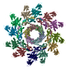 9lj8C M: map data used to model this data C: citing same article ( |
|---|---|
| Similar structure data |


-Links
 PDBj
PDBj- Assembly
| Deposited unit | 
|
|---|---|
| 1 |
|
-Components
-Protein , 7 types, 36 molecules 0stuvw123xyz456789GNUVWXghijkl...
| #1: Protein | Mass: 52085.984 Da / Num. of mol.: 6 / Source method: isolated from a natural source / Source: (natural) Escherichia phage Mu (virus) / References: UniProt: P08557#2: Protein | Mass: 53132.508 Da / Num. of mol.: 6 / Source method: isolated from a natural source / Source: (natural) Escherichia phage Mu (virus) / References: UniProt: P79678#3: Protein | Mass: 12840.470 Da / Num. of mol.: 6 / Source method: isolated from a natural source / Source: (natural) Escherichia phage Mu (virus) / References: UniProt: P79679#5: Protein | Mass: 41840.078 Da / Num. of mol.: 3 / Source method: isolated from a natural source / Source: (natural) Escherichia phage Mu (virus) / References: UniProt: P08558#7: Protein | Mass: 21720.525 Da / Num. of mol.: 3 / Source method: isolated from a natural source / Source: (natural) Escherichia phage Mu (virus) / References: UniProt: Q9T1V4#9: Protein | Mass: 73682.836 Da / Num. of mol.: 3 / Source method: isolated from a natural source / Source: (natural) Escherichia phage Mu (virus) / References: UniProt: Q9T1V6#10: Protein | Mass: 55412.551 Da / Num. of mol.: 9 / Source method: isolated from a natural source / Source: (natural) Escherichia phage Mu (virus) / References: UniProt: Q9T1V0 |
|---|
-Baseplate protein ... , 3 types, 24 molecules ABCDEFHIJKLMOPQRSTabcdef
| #4: Protein | Mass: 16340.159 Da / Num. of mol.: 6 / Source method: isolated from a natural source / Source: (natural) Escherichia phage Mu (virus) / References: UniProt: Q9T1V3#6: Protein | Mass: 38724.578 Da / Num. of mol.: 12 / Source method: isolated from a natural source / Source: (natural) Escherichia phage Mu (virus) / References: UniProt: Q9T1V2#8: Protein | Mass: 20489.059 Da / Num. of mol.: 6 / Source method: isolated from a natural source / Source: (natural) Escherichia phage Mu (virus) / References: UniProt: Q9T1V1 |
|---|
-Non-polymers , 2 types, 2 molecules 

| #11: Chemical | ChemComp-CA /  Mass: 40.078 Da / Num. of mol.: 1 / Source method: obtained synthetically / Formula: Ca / Feature type: SUBJECT OF INVESTIGATION Mass: 40.078 Da / Num. of mol.: 1 / Source method: obtained synthetically / Formula: Ca / Feature type: SUBJECT OF INVESTIGATION |
|---|---|
| #12: Chemical | ChemComp-FE /  Mass: 55.845 Da / Num. of mol.: 1 / Source method: obtained synthetically / Formula: Fe / Feature type: SUBJECT OF INVESTIGATION Mass: 55.845 Da / Num. of mol.: 1 / Source method: obtained synthetically / Formula: Fe / Feature type: SUBJECT OF INVESTIGATION |
-Details
| Has ligand of interest | Y |
|---|---|
| Has protein modification | N |
-Experimental details
-Experiment
| Experiment | Method: ELECTRON MICROSCOPY |
|---|---|
| EM experiment | Aggregation state: PARTICLE / 3D reconstruction method: single particle reconstruction |
- Sample preparation
| Component | Name: Escherichia phage Mu / Type: VIRUS / Entity ID: #1-#10 / Source: NATURAL |
|---|---|
| Source (natural) | Organism: Escherichia phage Mu (virus) |
| Details of virus | Empty: NO / Enveloped: NO / Isolate: SPECIES / Type: VIRION |
| Buffer solution | pH: 7.4 |
| Specimen | Embedding applied: NO / Shadowing applied: NO / Staining applied: NO / Vitrification applied: YES |
| Vitrification | Cryogen name: ETHANE |
- Electron microscopy imaging
| Experimental equipment |  Model: Titan Krios / Image courtesy: FEI Company |
|---|---|
| Microscopy | Model: TFS KRIOS |
| Electron gun | Electron source:  FIELD EMISSION GUN / Accelerating voltage: 300 kV / Illumination mode: FLOOD BEAM FIELD EMISSION GUN / Accelerating voltage: 300 kV / Illumination mode: FLOOD BEAM |
| Electron lens | Mode: BRIGHT FIELD / Nominal defocus max: 4000 nm / Nominal defocus min: 2000 nm |
| Image recording | Electron dose: 35 e/Å2 / Film or detector model: GATAN K3 (6k x 4k) |
- Processing
| CTF correction | Type: NONE |
|---|---|
| 3D reconstruction | Resolution: 3.3 Å / Resolution method: FSC 0.143 CUT-OFF / Num. of particles: 46000 / Symmetry type: POINT |
