 Movie
Movie Controller


+ Open data

- Basic information
| Entry |  | ||||||||||||
|---|---|---|---|---|---|---|---|---|---|---|---|---|---|
| Title | Neck structure of bacteriophage Mu in contracted state | ||||||||||||
 Map data Map data | |||||||||||||
 Sample Sample |
| ||||||||||||
 Keywords Keywords | portal / adaptor / phage / VIRAL PROTEIN | ||||||||||||
| Function / homology | Bacteriophage Mu, Gene product J / Bacteriophage Mu, Gp36 / Protein of unknown function DUF935 / Portal protein of Mu bacteriophage / symbiont genome ejection through host cell envelope, contractile tail mechanism / viral capsid / host cell cytoplasm / Gene product J / Portal protein Function and homology information Function and homology information | ||||||||||||
| Biological species |  Escherichia phage Mu (virus) Escherichia phage Mu (virus) | ||||||||||||
| Method | single particle reconstruction / cryo EM / Resolution: 3.6 Å | ||||||||||||
 Authors Authors | Liu HR / Zhou JQ | ||||||||||||
| Funding support |  China, 3 items China, 3 items
| ||||||||||||
 Citation Citation | Journal: J Virol / Year: 2025 Title: structures of the contractile nanomachine myophage Mu in both its extended and contracted states. Authors: Junquan Zhou / Liwen Wang / Hao Xiao / Wenyuan Chen / Zhonghua Liu / Jingdong Song / Jing Zheng / Hongrong Liu / Abstract: Myophage Mu is a representative of contractile nanomachines with a simple tail baseplate. It has the capacity to infect a range of intestinal bacteria and has extensive applications in genetic ...Myophage Mu is a representative of contractile nanomachines with a simple tail baseplate. It has the capacity to infect a range of intestinal bacteria and has extensive applications in genetic engineering research. Nevertheless, a comprehensive understanding of the entire structure and contractile mechanisms of Mu remains elusive. Using cryo-electron microscopy (cryo-EM), we resolved the asymmetric structures of Mu in both its extended and contracted states, the latter of which lacked the tail baseplate, at near-atomic resolutions. We built the atomic models for the extended Mu, encompassing the head, the connector complex, the tail, and the simple baseplate. It is noteworthy that we identified the position and structure of the tail tube initiator protein gp43 (referred to as the DNA circularization protein). The protein gp43 plays a crucial role not only in the baseplate assembly and DNA circularization but also in stabilizing the wedge-hub connection and mediating tail contraction. Except for the baseplate structure, the structural comparison of Mu in its extended and contracted states revealed that only the tail sheath protein gp39 and the C-terminus of the tail terminator protein gp37 undergo notable conformational changes to accommodate the tail contraction, whereas the remaining protein components remained unchanged. Our structures exhibited conserved properties among the majority of myophages, thereby providing valuable insights into the contraction mechanisms across myophages and contractile injection systems (CISs). IMPORTANCE: Despite extensive study, the asymmetric structures of phage Mu, a highly effective transposable myophage, remain unknown. In this study, we present the high-resolution structures of Mu in ...IMPORTANCE: Despite extensive study, the asymmetric structures of phage Mu, a highly effective transposable myophage, remain unknown. In this study, we present the high-resolution structures of Mu in both its extended and contracted states. The comparison of the two structures allows for the illustration of detailed conformational changes of the head-to-tail complex during the process of tail contraction. The contraction mechanism of Mu is highly conserved and widely adapted to all contractile nanomachines that share common structural features with Mu. | ||||||||||||
| History |
|
- Structure visualization
| Supplemental images |
|---|

- Downloads & links
-EMDB archive
| Map data | emd_62462.map.gz | 164.8 MB | EMDB map data format | |
|---|---|---|---|---|
| Header (meta data) | emd-62462-v30.xmlemd-62462.xml | 17 KB 17 KB | Display Display | EMDB header |
| Images |  emd_62462.png emd_62462.png | 63.6 KB | ||
| Filedesc metadata | emd-62462.cif.gz | 5.9 KB | ||
| Others | emd_62462_half_map_1.map.gzemd_62462_half_map_2.map.gz | 164.6 MB 164.6 MB | ||
| Archive directory |  http://ftp.pdbj.org/pub/emdb/structures/EMD-62462ftp://ftp.pdbj.org/pub/emdb/structures/EMD-62462 http://ftp.pdbj.org/pub/emdb/structures/EMD-62462ftp://ftp.pdbj.org/pub/emdb/structures/EMD-62462 | HTTPS FTP |
-Related structure data
| Related structure data |  9knuMC  9jodC  9khxC  9khyC  9ki1C 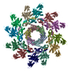 9lj8C M: atomic model generated by this map C: citing same article ( |
|---|---|
| Similar structure data |


-Links
| EMDB pages | EMDB (EBI/PDBe) / EMDataResource |
|---|
-Map
| File | Download / File: emd_62462.map.gz / Format: CCP4 / Size: 178 MB / Type: IMAGE STORED AS FLOATING POINT NUMBER (4 BYTES) | ||||||||||||||||||||||||||||||||||||
|---|---|---|---|---|---|---|---|---|---|---|---|---|---|---|---|---|---|---|---|---|---|---|---|---|---|---|---|---|---|---|---|---|---|---|---|---|---|
| Projections & slices | Image control
Images are generated by Spider. | ||||||||||||||||||||||||||||||||||||
| Voxel size | X=Y=Z: 1.06 Å | ||||||||||||||||||||||||||||||||||||
| Density |
| ||||||||||||||||||||||||||||||||||||
| Symmetry | Space group: 1 | ||||||||||||||||||||||||||||||||||||
| Details | EMDB XML:
|
 Z (Sec.)
 Y (Row.)
 X (Col.)


-Supplemental data
-Half map: #2
| File | emd_62462_half_map_1.map | ||||||||||||
|---|---|---|---|---|---|---|---|---|---|---|---|---|---|
| Projections & Slices |
| ||||||||||||
| Density Histograms |


-Half map: #1
| File | emd_62462_half_map_2.map | ||||||||||||
|---|---|---|---|---|---|---|---|---|---|---|---|---|---|
| Projections & Slices |
| ||||||||||||
| Density Histograms |


- Sample components
-Entire : Escherichia phage Mu
| Entire | Name: Escherichia phage Mu (virus) |
|---|---|
| Components |
|
-Supramolecule #1: Escherichia phage Mu
| Supramolecule | Name: Escherichia phage Mu / type: complex / ID: 1 / Parent: 0 / Macromolecule list: all |
|---|---|
| Source (natural) | Organism: Escherichia phage Mu (virus) |
-Macromolecule #1: Portal protein
| Macromolecule | Name: Portal protein / type: protein_or_peptide / ID: 1 / Number of copies: 12 / Enantiomer: LEVO |
|---|---|
| Source (natural) | Organism: Escherichia phage Mu (virus) |
| Molecular weight | Theoretical: 56.952305 KDa |
| Sequence | String: MGRILDISGQ PFDFDDEMQS RSDELAMVMK RTQEHPSSGV TPNRAAQMLR DAERGDLTAQ ADLAFDMEEK DTHLFSELSK RRLAIQALE WRIAPARDAS AQEKKDADML NEYLHDAAWF EDALFDAGDA ILKGYSMQEI EWGWLGKMRV PVALHHRDPA L FCANPDNL ...String: MGRILDISGQ PFDFDDEMQS RSDELAMVMK RTQEHPSSGV TPNRAAQMLR DAERGDLTAQ ADLAFDMEEK DTHLFSELSK RRLAIQALE WRIAPARDAS AQEKKDADML NEYLHDAAWF EDALFDAGDA ILKGYSMQEI EWGWLGKMRV PVALHHRDPA L FCANPDNL NELRLRDASY HGLELQPFGW FMHRAKSRTG YVGTNGLVRT LIWPFIFKNY SVRDFAEFLE IYGLPMRVGK YP TGSTNRE KATLMQAVMD IGRRAGGIIP MGMTLDFQSA ADGQSDPFMA MIGWAEKAIS KAILGGTLTT EAGDKGARSL GEV HDEVRR EIRNADVGQL ARSINRDLIY PLLALNSDST IDINRLPGIV FDTSEAGDIT ALSDAIPKLA AGMRIPVSWI QEKL HIPQP VGDEAVFTIQ PVVPDNGSQK EAALSAEDIP QEDDIDRMGV SPEDWQRSVD PLLKPVIFSV LKDGPEAAMN KAASL YPQM DDAELIDMLT RAIFVADIWG RLDAAADH UniProtKB: Portal protein |
-Macromolecule #2: Gene product J
| Macromolecule | Name: Gene product J / type: protein_or_peptide / ID: 2 / Number of copies: 12 / Enantiomer: LEVO |
|---|---|
| Source (natural) | Organism: Escherichia phage Mu (virus) |
| Molecular weight | Theoretical: 15.742675 KDa |
| Sequence | String: MNYATVNDLC ARYTRTRLDI LTRPKTADGQ PDDAVAEQAL ADASAFIDGY LAARFVLPLT VVPSLLKRQC CVVAWFYLNE SQPTEQITA TYRDTVRWLE QVRDGKTDPG VESRTAASPE GEDLVQVQSD PPVFSRKQKG FI UniProtKB: Gene product J |
-Experimental details
-Structure determination
| Method | cryo EM |
|---|---|
 Processing Processing | single particle reconstruction |
| Aggregation state | particle |
-Sample preparation
| Buffer | pH: 7.4 |
|---|---|
| Vitrification | Cryogen name: ETHANE |
- Electron microscopy
| Microscope | TFS KRIOS |
|---|---|
| Image recording | Film or detector model: GATAN K3 (6k x 4k) / Average electron dose: 35.0 e/Å2 |
| Electron beam | Acceleration voltage: 300 kV / Electron source:  FIELD EMISSION GUN FIELD EMISSION GUN |
| Electron optics | Illumination mode: FLOOD BEAM / Imaging mode: BRIGHT FIELD / Nominal defocus max: 4.0 µm / Nominal defocus min: 2.0 µm |
| Experimental equipment |  Model: Titan Krios / Image courtesy: FEI Company |
