Movie
Movie Controller


+ Open data

- Basic information
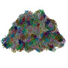
| Entry | Database: PDB / ID: 9k9w | |||||||||
|---|---|---|---|---|---|---|---|---|---|---|
| Title | structure of bundle-shaped PBS with short rod | |||||||||
 Components Components |
| |||||||||
 Keywords Keywords | PHOTOSYNTHESIS / phycobilisome / light-harvesting complex | |||||||||
| Function / homology |  Function and homology information Function and homology informationphycobilisome / plasma membrane-derived thylakoid membrane / photosynthesis / lyase activity Similarity search - Function | |||||||||
| Biological species |  Gloeobacter violaceus PCC 7421 (bacteria) Gloeobacter violaceus PCC 7421 (bacteria) | |||||||||
| Method | ELECTRON MICROSCOPY / single particle reconstruction / cryo EM / Resolution: 2.8 Å | |||||||||
 Authors Authors | Sui, S.-F. / Ma, J. / You, X. / Sun, S. | |||||||||
| Funding support |  China, 2items China, 2items
| |||||||||
 Citation Citation | Journal: Nat Commun / Year: 2025 Title: Light-induced structural adaptation of the bundle-shaped phycobilisome from thylakoid-lacking cyanobacterium Gloeobacter violaceus. Authors: Jianfei Ma / Xin You / Shan Sun / Sen-Fang Sui / Abstract: Gloeobacter diverged from other lineages early in cyanobacterial evolution, preferentially growing under low light intensity conditions. Among cyanobacteria, G. violaceus exhibits unique features, ...Gloeobacter diverged from other lineages early in cyanobacterial evolution, preferentially growing under low light intensity conditions. Among cyanobacteria, G. violaceus exhibits unique features, including lack of a thylakoid membrane and bundle-shaped antenna phycobilisomes (PBSs), densely packed and well-organized on the plasma membrane. However, without high-resolution structures, it has remained unclear how G. violaceus PBSs assemble into a bundle-shaped configuration. Here we solve the cryo-EM structures of PBSs from G. violaceus cells cultured under low (Sr-PBS) or moderate (Lr-PBS) light intensity. These structures reveal two unique linker proteins, L and L, that play a key role in the PBS architecture. Analysis of the bilin arrangement indicates that the bundle-shaped structure allows efficient energy transfer among rods. Moreover, comparison between Lr-PBS and Sr-PBS uncovers a distinct mode of adaption to increased light intensity wherein the ApcA-ApcB-ApcD layer can be blocked from binding to the core by altering structural elements exclusively found in the G. violaceus L. This study illustrates previously unrecognized mechanisms of assembly and adaptation to varying light intensity in the bundle-shaped PBS of G. violaceus. | |||||||||
| History |
|
- Structure visualization
| Structure viewer | Molecule: MolmilJmol/JSmol |
|---|
- Downloads & links
-Download
| PDBx/mmCIF format | 9k9w.cif.gz | 8.6 MB | Display | PDBx/mmCIF format |
|---|---|---|---|---|
| PDB format | pdb9k9w.ent.gz | Display | PDB format | |
| PDBx/mmJSON format | 9k9w.json.gz | Tree view | PDBx/mmJSON format | |
| Others |  Other downloads Other downloads |
-Validation report
| Arichive directory | https://data.pdbj.org/pub/pdb/validation_reports/k9/9k9wftp://data.pdbj.org/pub/pdb/validation_reports/k9/9k9w | HTTPS FTP |
|---|
-Related structure data
| Related structure data |  62201MC  9m3jC  36381 36382 8jkx M: map data used to model this data C: citing same article ( |
|---|---|
| Similar structure data |


-Links
 PDBj
PDBj- Assembly
| Deposited unit | 
|
|---|---|
| 1 |
|
-Components
-Allophycocyanin ... , 2 types, 82 molecules AZL5N5B5D5F5H5J5d5f5P5R5T5V5X5Z5b5P7R7B7D7F7H7J7L7N7h7j7T7...
| #1: Protein | Mass: 17241.664 Da / Num. of mol.: 42 / Source method: isolated from a natural source / Source: (natural) Gloeobacter violaceus PCC 7421 (bacteria) / References: UniProt: Q7NL79#8: Protein | Mass: 17539.039 Da / Num. of mol.: 40 / Source method: isolated from a natural source / Source: (natural) Gloeobacter violaceus PCC 7421 (bacteria) / References: UniProt: Q7NL80 |
|---|
-Protein , 7 types, 182 molecules AACAEAGAIAKAOAQASAUAWAYAO1Q1S1A1G3I3K3G2I2K2A3C3E3A4C4O3Q3S3...
| #2: Protein | Mass: 17679.852 Da / Num. of mol.: 84 / Source method: isolated from a natural source / Source: (natural) Gloeobacter violaceus PCC 7421 (bacteria) / References: UniProt: Q7M7F7#3: Protein | Mass: 18478.953 Da / Num. of mol.: 84 / Source method: isolated from a natural source / Source: (natural) Gloeobacter violaceus PCC 7421 (bacteria) / References: UniProt: Q7M7C7#6: Protein | Mass: 81544.312 Da / Num. of mol.: 2 / Source method: isolated from a natural source / Source: (natural) Gloeobacter violaceus PCC 7421 (bacteria) / References: UniProt: Q7NGT2#7: Protein | Mass: 7776.872 Da / Num. of mol.: 2 / Source method: isolated from a natural source / Source: (natural) Gloeobacter violaceus PCC 7421 (bacteria) / References: UniProt: Q7NL59#9: Protein | Mass: 7633.809 Da / Num. of mol.: 6 / Source method: isolated from a natural source / Source: (natural) Gloeobacter violaceus PCC 7421 (bacteria) / References: UniProt: Q7NL78#10: Protein | Mass: 130002.258 Da / Num. of mol.: 2 / Source method: isolated from a natural source / Source: (natural) Gloeobacter violaceus PCC 7421 (bacteria) / References: UniProt: Q7NL81#11: Protein | Mass: 92127.602 Da / Num. of mol.: 2 / Source method: isolated from a natural source / Source: (natural) Gloeobacter violaceus PCC 7421 (bacteria) / References: UniProt: Q7NL64 |
|---|
-Phycocyanin-associated rod linker ... , 2 types, 12 molecules MAM3M4M8M9M1NAN3Z4N9Z8N1
| #4: Protein | Mass: 30913.822 Da / Num. of mol.: 6 / Source method: isolated from a natural source Details: There is no expression tag in the Phycocyanin-associated rod linker proteins (MA, M3, M4, M8, M9 and M1) or the Phycocyanin-associated rod linker proteins (NA, N3, Z4, N9, Z8 and N1). We ...Details: There is no expression tag in the Phycocyanin-associated rod linker proteins (MA, M3, M4, M8, M9 and M1) or the Phycocyanin-associated rod linker proteins (NA, N3, Z4, N9, Z8 and N1). We align all amino acids by their electron density map. Source: (natural) Gloeobacter violaceus PCC 7421 (bacteria) / References: UniProt: Q7NGF2#5: Protein | Mass: 31085.066 Da / Num. of mol.: 6 / Source method: isolated from a natural source / Source: (natural) Gloeobacter violaceus PCC 7421 (bacteria) / References: UniProt: Q7NM19 |
|---|
-Non-polymers , 1 types, 336 molecules 
| #12: Chemical | ChemComp-CYC /  Mass: 588.694 Da / Num. of mol.: 336 / Source method: obtained synthetically / Formula: C33H40N4O6 / Feature type: SUBJECT OF INVESTIGATION Mass: 588.694 Da / Num. of mol.: 336 / Source method: obtained synthetically / Formula: C33H40N4O6 / Feature type: SUBJECT OF INVESTIGATION |
|---|
-Details
| Has ligand of interest | Y |
|---|---|
| Has protein modification | Y |
-Experimental details
-Experiment
| Experiment | Method: ELECTRON MICROSCOPY |
|---|---|
| EM experiment | Aggregation state: PARTICLE / 3D reconstruction method: single particle reconstruction |
- Sample preparation
| Component | Name: phycobilisome / Type: COMPLEX / Entity ID: #6, #5 / Source: NATURAL | |||||||||||||||
|---|---|---|---|---|---|---|---|---|---|---|---|---|---|---|---|---|
| Molecular weight | Value: 14700 kDa/nm / Experimental value: NO | |||||||||||||||
| Source (natural) | Organism:  Porphyridium purpureum (eukaryote) Porphyridium purpureum (eukaryote) | |||||||||||||||
| Buffer solution | pH: 7 | |||||||||||||||
| Buffer component |
| |||||||||||||||
| Specimen | Embedding applied: NO / Shadowing applied: NO / Staining applied: NO / Vitrification applied: YES | |||||||||||||||
| Specimen support | Grid material: COPPER / Grid mesh size: 300 divisions/in. / Grid type: Quantifoil R2/1 | |||||||||||||||
| Vitrification | Instrument: FEI VITROBOT MARK IV / Cryogen name: ETHANE / Humidity: 100 % / Chamber temperature: 18 K |
- Electron microscopy imaging
| Experimental equipment |  Model: Tecnai F30 / Image courtesy: FEI Company |
|---|---|
| Microscopy | Model: FEI TECNAI F30 |
| Electron gun | Electron source:  FIELD EMISSION GUN / Accelerating voltage: 300 kV / Illumination mode: FLOOD BEAM FIELD EMISSION GUN / Accelerating voltage: 300 kV / Illumination mode: FLOOD BEAM |
| Electron lens | Mode: BRIGHT FIELD / Nominal magnification: 64000 X / Calibrated magnification: 64000 X / Nominal defocus max: 2300 nm / Nominal defocus min: 1300 nm / Calibrated defocus min: 1300 nm / Calibrated defocus max: 2300 nm / Cs: 0.001 mm / C2 aperture diameter: 100 µm / Alignment procedure: COMA FREE |
| Specimen holder | Cryogen: NITROGEN / Specimen holder model: FEI TITAN KRIOS AUTOGRID HOLDER / Temperature (max): 77 K / Temperature (min): 77 K |
| Image recording | Average exposure time: 8.2 sec. / Electron dose: 50 e/Å2 / Detector mode: SUPER-RESOLUTION / Film or detector model: GATAN K2 SUMMIT (4k x 4k) / Num. of grids imaged: 2 / Num. of real images: 6438 |
| EM imaging optics | Energyfilter name: GIF Quantum ER / Energyfilter slit width: 20 eV Spherical aberration corrector: Spherical aberration (Cs) corrector |
| Image scans | Width: 4096 / Height: 4096 / Movie frames/image: 32 / Used frames/image: 1-32 |
- Processing
| EM software |
| ||||||||||||||||||||||||||||||||||||
|---|---|---|---|---|---|---|---|---|---|---|---|---|---|---|---|---|---|---|---|---|---|---|---|---|---|---|---|---|---|---|---|---|---|---|---|---|---|
| CTF correction | Type: NONE | ||||||||||||||||||||||||||||||||||||
| Particle selection | Num. of particles selected: 1042622 | ||||||||||||||||||||||||||||||||||||
| Symmetry | Point symmetry: C2 (2 fold cyclic) | ||||||||||||||||||||||||||||||||||||
| 3D reconstruction | Resolution: 2.8 Å / Resolution method: FSC 0.143 CUT-OFF / Num. of particles: 389430 / Symmetry type: POINT | ||||||||||||||||||||||||||||||||||||
| Atomic model building | B value: 62.05 / Protocol: RIGID BODY FIT / Space: REAL / Target criteria: 0,99 | ||||||||||||||||||||||||||||||||||||
| Atomic model building | PDB-ID: 6KGX Accession code: 6KGX / Source name: PDB / Type: experimental model |