Movie
Movie Controller


+ Open data

- Basic information
| Entry |  | |||||||||
|---|---|---|---|---|---|---|---|---|---|---|
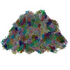
| Title | structure of bundle-shaped PBS with short rod | |||||||||
 Map data Map data | structure of bundle-shaped PBS with short rod at 2.80A | |||||||||
 Sample Sample |
| |||||||||
 Keywords Keywords | phycobilisome / light-harvesting complex / photosynthesis | |||||||||
| Function / homology |  Function and homology information Function and homology informationphycobilisome / plasma membrane-derived thylakoid membrane / photosynthesis / lyase activity Similarity search - Function | |||||||||
| Biological species |  Porphyridium purpureum (eukaryote) / Porphyridium purpureum (eukaryote) /  Gloeobacter violaceus PCC 7421 (bacteria) Gloeobacter violaceus PCC 7421 (bacteria) | |||||||||
| Method | single particle reconstruction / cryo EM / Resolution: 2.8 Å | |||||||||
 Authors Authors | Sui S-F / Ma J / You X / Sun S | |||||||||
| Funding support |  China, 2 items China, 2 items
| |||||||||
 Citation Citation | Journal: Nat Commun / Year: 2025 Title: Light-induced structural adaptation of the bundle-shaped phycobilisome from thylakoid-lacking cyanobacterium Gloeobacter violaceus. Authors: Jianfei Ma / Xin You / Shan Sun / Sen-Fang Sui / Abstract: Gloeobacter diverged from other lineages early in cyanobacterial evolution, preferentially growing under low light intensity conditions. Among cyanobacteria, G. violaceus exhibits unique features, ...Gloeobacter diverged from other lineages early in cyanobacterial evolution, preferentially growing under low light intensity conditions. Among cyanobacteria, G. violaceus exhibits unique features, including lack of a thylakoid membrane and bundle-shaped antenna phycobilisomes (PBSs), densely packed and well-organized on the plasma membrane. However, without high-resolution structures, it has remained unclear how G. violaceus PBSs assemble into a bundle-shaped configuration. Here we solve the cryo-EM structures of PBSs from G. violaceus cells cultured under low (Sr-PBS) or moderate (Lr-PBS) light intensity. These structures reveal two unique linker proteins, L and L, that play a key role in the PBS architecture. Analysis of the bilin arrangement indicates that the bundle-shaped structure allows efficient energy transfer among rods. Moreover, comparison between Lr-PBS and Sr-PBS uncovers a distinct mode of adaption to increased light intensity wherein the ApcA-ApcB-ApcD layer can be blocked from binding to the core by altering structural elements exclusively found in the G. violaceus L. This study illustrates previously unrecognized mechanisms of assembly and adaptation to varying light intensity in the bundle-shaped PBS of G. violaceus. | |||||||||
| History |
|
- Structure visualization
| Supplemental images |
|---|

- Downloads & links
-EMDB archive
| Map data | emd_62201.map.gz | 632.2 MB | EMDB map data format | |
|---|---|---|---|---|
| Header (meta data) | emd-62201-v30.xmlemd-62201.xml | 35.7 KB 35.7 KB | Display Display | EMDB header |
| Images |  emd_62201.png emd_62201.png | 154.6 KB | ||
| Filedesc metadata | emd-62201.cif.gz | 9.2 KB | ||
| Others | emd_62201_half_map_1.map.gzemd_62201_half_map_2.map.gz | 606.7 MB 621.5 MB | ||
| Archive directory |  http://ftp.pdbj.org/pub/emdb/structures/EMD-62201ftp://ftp.pdbj.org/pub/emdb/structures/EMD-62201 http://ftp.pdbj.org/pub/emdb/structures/EMD-62201ftp://ftp.pdbj.org/pub/emdb/structures/EMD-62201 | HTTPS FTP |
-Related structure data
| Related structure data |  9k9wMC  9m3jC  36381 36382 M: atomic model generated by this map C: citing same article ( |
|---|---|
| Similar structure data |


-Links
| EMDB pages | EMDB (EBI/PDBe) / EMDataResource |
|---|
-Map
| File | Download / File: emd_62201.map.gz / Format: CCP4 / Size: 669.9 MB / Type: IMAGE STORED AS FLOATING POINT NUMBER (4 BYTES) | ||||||||||||||||||||||||||||||||||||
|---|---|---|---|---|---|---|---|---|---|---|---|---|---|---|---|---|---|---|---|---|---|---|---|---|---|---|---|---|---|---|---|---|---|---|---|---|---|
| Annotation | structure of bundle-shaped PBS with short rod at 2.80A | ||||||||||||||||||||||||||||||||||||
| Projections & slices | Image control
Images are generated by Spider. | ||||||||||||||||||||||||||||||||||||
| Voxel size | X=Y=Z: 1.0979 Å | ||||||||||||||||||||||||||||||||||||
| Density |
| ||||||||||||||||||||||||||||||||||||
| Symmetry | Space group: 1 | ||||||||||||||||||||||||||||||||||||
| Details | EMDB XML:
|
 Z (Sec.)
 Y (Row.)
 X (Col.)


-Supplemental data
-Half map: structure of bundle-shaped PBS with short rod at 2.80A halfB
| File | emd_62201_half_map_1.map | ||||||||||||
|---|---|---|---|---|---|---|---|---|---|---|---|---|---|
| Annotation | structure of bundle-shaped PBS with short rod at 2.80A halfB | ||||||||||||
| Projections & Slices |
| ||||||||||||
| Density Histograms |


-Half map: structure of bundle-shaped PBS with short rod at 2.80A halfA
| File | emd_62201_half_map_2.map | ||||||||||||
|---|---|---|---|---|---|---|---|---|---|---|---|---|---|
| Annotation | structure of bundle-shaped PBS with short rod at 2.80A halfA | ||||||||||||
| Projections & Slices |
| ||||||||||||
| Density Histograms |


- Sample components
+Entire : phycobilisome
+Supramolecule #1: phycobilisome
+Macromolecule #1: Allophycocyanin beta subunit
+Macromolecule #2: Phycocyanin alpha chain
+Macromolecule #3: Phycocyanin beta chain
+Macromolecule #4: Phycocyanin-associated rod linker protein
+Macromolecule #5: Phycocyanin-associated rod linker protein
+Macromolecule #6: Glr2806 protein
+Macromolecule #7: CpcD protein
+Macromolecule #8: Allophycocyanin alpha subunit
+Macromolecule #9: Phycobilisome 7.8 kDa linker polypeptide, allophycocyanin-associa...
+Macromolecule #10: Phycobiliprotein ApcE
+Macromolecule #11: Glr1262 protein
+Macromolecule #12: PHYCOCYANOBILIN

-Experimental details
-Structure determination
| Method | cryo EM |
|---|---|
 Processing Processing | single particle reconstruction |
| Aggregation state | particle |
-Sample preparation
| Buffer | pH: 7 Component:
| |||||||||
|---|---|---|---|---|---|---|---|---|---|---|
| Grid | Model: Quantifoil R2/1 / Material: COPPER / Mesh: 300 / Support film - Material: CARBON / Support film - topology: CONTINUOUS / Support film - Film thickness: 2 | |||||||||
| Vitrification | Cryogen name: ETHANE / Chamber humidity: 100 % / Chamber temperature: 18 K / Instrument: FEI VITROBOT MARK IV |
- Electron microscopy
| Microscope | FEI TECNAI F30 |
|---|---|
| Temperature | Min: 77.0 K / Max: 77.0 K |
| Specialist optics | Spherical aberration corrector: Spherical aberration (Cs) corrector Energy filter - Name: GIF Quantum ER / Energy filter - Slit width: 20 eV |
| Image recording | Film or detector model: GATAN K2 SUMMIT (4k x 4k) / Detector mode: SUPER-RESOLUTION / Digitization - Dimensions - Width: 4096 pixel / Digitization - Dimensions - Height: 4096 pixel / Digitization - Frames/image: 1-32 / Number grids imaged: 2 / Number real images: 6438 / Average exposure time: 8.2 sec. / Average electron dose: 50.0 e/Å2 |
| Electron beam | Acceleration voltage: 300 kV / Electron source:  FIELD EMISSION GUN FIELD EMISSION GUN |
| Electron optics | C2 aperture diameter: 100.0 µm / Calibrated defocus max: 2.3000000000000003 µm / Calibrated defocus min: 1.3 µm / Calibrated magnification: 64000 / Illumination mode: FLOOD BEAM / Imaging mode: BRIGHT FIELD / Cs: 0.001 mm / Nominal defocus max: 2.3000000000000003 µm / Nominal defocus min: 1.3 µm / Nominal magnification: 64000 |
| Sample stage | Specimen holder model: FEI TITAN KRIOS AUTOGRID HOLDER / Cooling holder cryogen: NITROGEN |
| Experimental equipment |  Model: Tecnai F30 / Image courtesy: FEI Company |