 Movie
Movie Controller


+ Open data

- Basic information
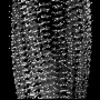
| Entry | Database: PDB / ID: 9jsu | |||||||||
|---|---|---|---|---|---|---|---|---|---|---|
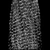
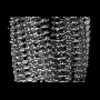
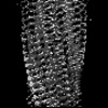
| Title | Wild-type native PMEL amyloid - polymorph 2 | |||||||||
 Components Components | M-alpha | |||||||||
 Keywords Keywords | STRUCTURAL PROTEIN / melanosome / melanoma / pigment / melanin / amyloid / glaucoma | |||||||||
| Function / homology |  Function and homology information Function and homology informationcis-Golgi network membrane / positive regulation of melanin biosynthetic process / melanin biosynthetic process / melanosome membrane / melanosome organization / multivesicular body, internal vesicle / multivesicular body membrane / Regulation of MITF-M-dependent genes involved in pigmentation / melanosome / endoplasmic reticulum membrane ...cis-Golgi network membrane / positive regulation of melanin biosynthetic process / melanin biosynthetic process / melanosome membrane / melanosome organization / multivesicular body, internal vesicle / multivesicular body membrane / Regulation of MITF-M-dependent genes involved in pigmentation / melanosome / endoplasmic reticulum membrane / endoplasmic reticulum / Golgi apparatus / extracellular exosome / identical protein binding / plasma membrane Similarity search - Function | |||||||||
| Biological species |  Homo sapiens (human) Homo sapiens (human) | |||||||||
| Method | ELECTRON MICROSCOPY / single particle reconstruction / cryo EM / Resolution: 1.79 Å | |||||||||
 Authors Authors | Oda, T. / Yanagisawa, H. | |||||||||
| Funding support |  Japan, Japan,  France, 2items France, 2items
| |||||||||
 Citation Citation | Journal: Nat Commun / Year: 2025 Title: Cryo-EM of wild-type and mutant PMEL amyloid cores reveals structural mechanism of pigment dispersion syndrome. Authors: Haruaki Yanagisawa / Harumi Arai / Tony Wang / Hideyuki Miyazawa / Masahide Kikkawa / Toshiyuki Oda / Abstract: PMEL amyloids serve as essential scaffolds for melanin deposition in melanosomes, playing a crucial role in pigmentation. Despite their importance, the high-resolution structure of PMEL amyloids has ...PMEL amyloids serve as essential scaffolds for melanin deposition in melanosomes, playing a crucial role in pigmentation. Despite their importance, the high-resolution structure of PMEL amyloids has remained unresolved. Using cryo-electron microscopy, we determine near-atomic resolution structures of wild-type PMEL amyloid core, revealing two distinct polymorphic forms with structural features. We further investigate the pathogenic G175S mutation associated with pigment dispersion syndrome (PDS). Structural analysis reveales that G175S introduces an additional hydrogen bond, stabilizing an alternative fibril conformation. In vitro, the G175S mutant exhibits a fourfold increase in polymerization efficiency compared to the wild type. In cells, G175S expression resultes in a twofold increase in intracellular amyloid content and a ~70% increase in extracellular amyloids, without altering melanosome morphology or number. These results indicate that the G175S mutation enhances amyloidogenesis within melanosomes, elevating amyloid load and potentially contributing to PDS pathophysiology. This study provides molecular insights into PMEL amyloid formation, highlighting its structural diversity and dysregulation in pigmentation disorders. | |||||||||
| History |
|
- Structure visualization
| Structure viewer | Molecule: MolmilJmol/JSmol |
|---|
- Downloads & links
-Download
| PDBx/mmCIF format | 9jsu.cif.gz | 89 KB | Display | PDBx/mmCIF format |
|---|---|---|---|---|
| PDB format | pdb9jsu.ent.gz | 69 KB | Display | PDB format |
| PDBx/mmJSON format | 9jsu.json.gz | Tree view | PDBx/mmJSON format | |
| Others |  Other downloads Other downloads |
-Validation report
| Summary document | 9jsu_validation.pdf.gz | 1.2 MB | Display | wwPDB validaton report |
|---|---|---|---|---|
| Full document | 9jsu_full_validation.pdf.gz | 1.2 MB | Display | |
| Data in XML | 9jsu_validation.xml.gz | 19.6 KB | Display | |
| Data in CIF | 9jsu_validation.cif.gz | 29 KB | Display | |
| Arichive directory | https://data.pdbj.org/pub/pdb/validation_reports/js/9jsuftp://data.pdbj.org/pub/pdb/validation_reports/js/9jsu | HTTPS FTP |
-Related structure data
| Related structure data | 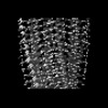 61783MC  9jstC  9jsvC  9jswC 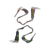 9jsxC C: citing same article ( M: map data used to model this data |
|---|---|
| Similar structure data |
-Links
 PDBj
PDBj
- Assembly
| Deposited unit | 
|
|---|---|
| 1 |
|
-Components
| #1: Protein/peptide | Mass: 3611.115 Da / Num. of mol.: 8 / Source method: isolated from a natural source / Source: (natural) Homo sapiens (human) / Cell line: HMV-II / References: UniProt: P40967Has protein modification | N | |
|---|
-Experimental details
-Experiment
| Experiment | Method: ELECTRON MICROSCOPY |
|---|---|
| EM experiment | Aggregation state: FILAMENT / 3D reconstruction method: single particle reconstruction |
- Sample preparation
| Component | Name: Wild-type native PMEL amyloid extracted from melanoma cell line Type: CELL / Entity ID: all / Source: NATURAL |
|---|---|
| Source (natural) | Organism: Homo sapiens (human) / Organelle: Melanosome |
| Buffer solution | pH: 4.4 |
| Buffer component | Conc.: 150 mM / Name: Sodium acetate / Formula: CH3COONa |
| Specimen | Conc.: 0.3 mg/ml / Embedding applied: NO / Shadowing applied: NO / Staining applied: NO / Vitrification applied: YES / Details: This sample was monodisperse. |
| Specimen support | Grid material: GOLD / Grid mesh size: 300 divisions/in. / Grid type: UltrAuFoil R1.2/1.3 |
| Vitrification | Instrument: FEI VITROBOT MARK IV / Cryogen name: ETHANE / Humidity: 100 % / Chamber temperature: 283 K |
- Electron microscopy imaging
| Microscopy | Model: JEOL CRYO ARM 300 |
|---|---|
| Electron gun | Electron source:  FIELD EMISSION GUN / Accelerating voltage: 300 kV / Illumination mode: FLOOD BEAM FIELD EMISSION GUN / Accelerating voltage: 300 kV / Illumination mode: FLOOD BEAM |
| Electron lens | Mode: BRIGHT FIELD / Nominal magnification: 60000 X / Nominal defocus max: 2000 nm / Nominal defocus min: 500 nm / Cs: 2.7 mm / Alignment procedure: COMA FREE |
| Specimen holder | Cryogen: NITROGEN |
| Image recording | Average exposure time: 5.5 sec. / Electron dose: 50 e/Å2 / Film or detector model: GATAN K3 (6k x 4k) |
| EM imaging optics | Energyfilter name: In-column Omega Filter / Energyfilter slit width: 20 eV |
- Processing
| CTF correction | Type: PHASE FLIPPING AND AMPLITUDE CORRECTION |
|---|---|
| 3D reconstruction | Resolution: 1.79 Å / Resolution method: FSC 0.143 CUT-OFF / Num. of particles: 352878 / Symmetry type: POINT |
