 Movie
Movie Controller


+ Open data

- Basic information
| Entry | Database: PDB / ID: 9jr3 | |||||||||||||||||||||||||||
|---|---|---|---|---|---|---|---|---|---|---|---|---|---|---|---|---|---|---|---|---|---|---|---|---|---|---|---|---|
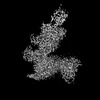
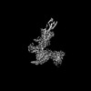
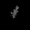
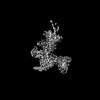
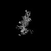
| Title | Cryo-EM structure of PTH-PTH1R-Gq (tilted state) | |||||||||||||||||||||||||||
 Components Components |
| |||||||||||||||||||||||||||
 Keywords Keywords | SIGNALING PROTEIN/IMMUNE SYSTEM / Class B GPCRs / Gq / PTH / SIGNALING PROTEIN / SIGNALING PROTEIN-IMMUNE SYSTEM complex | |||||||||||||||||||||||||||
| Function / homology |  Function and homology information Function and homology informationmacromolecule biosynthetic process / parathyroid hormone receptor binding / type 1 parathyroid hormone receptor binding / negative regulation of bone mineralization involved in bone maturation / negative regulation of apoptotic process in bone marrow cell / positive regulation of osteoclast proliferation / response to parathyroid hormone / positive regulation of cell proliferation in bone marrow / hormone-mediated apoptotic signaling pathway / adenylate cyclase-activating G protein-coupled cAMP receptor signaling pathway ...macromolecule biosynthetic process / parathyroid hormone receptor binding / type 1 parathyroid hormone receptor binding / negative regulation of bone mineralization involved in bone maturation / negative regulation of apoptotic process in bone marrow cell / positive regulation of osteoclast proliferation / response to parathyroid hormone / positive regulation of cell proliferation in bone marrow / hormone-mediated apoptotic signaling pathway / adenylate cyclase-activating G protein-coupled cAMP receptor signaling pathway / parathyroid hormone receptor activity / positive regulation of signal transduction / magnesium ion homeostasis / response to fibroblast growth factor / cAMP metabolic process / phosphate ion homeostasis / G-protein activation / Activation of the phototransduction cascade / Glucagon-type ligand receptors / Thromboxane signalling through TP receptor / Sensory perception of sweet, bitter, and umami (glutamate) taste / G beta:gamma signalling through PI3Kgamma / G beta:gamma signalling through CDC42 / Cooperation of PDCL (PhLP1) and TRiC/CCT in G-protein beta folding / Activation of G protein gated Potassium channels / Inhibition of voltage gated Ca2+ channels via Gbeta/gamma subunits / Ca2+ pathway / G alpha (z) signalling events / High laminar flow shear stress activates signaling by PIEZO1 and PECAM1:CDH5:KDR in endothelial cells / Glucagon-like Peptide-1 (GLP1) regulates insulin secretion / G protein-coupled peptide receptor activity / Class B/2 (Secretin family receptors) / Vasopressin regulates renal water homeostasis via Aquaporins / negative regulation of chondrocyte differentiation / Adrenaline,noradrenaline inhibits insulin secretion / ADP signalling through P2Y purinoceptor 12 / G alpha (q) signalling events / osteoblast development / response to vitamin D / G alpha (i) signalling events / Thrombin signalling through proteinase activated receptors (PARs) / Activation of G protein gated Potassium channels / G-protein activation / G beta:gamma signalling through PI3Kgamma / Prostacyclin signalling through prostacyclin receptor / G beta:gamma signalling through PLC beta / ADP signalling through P2Y purinoceptor 1 / Thromboxane signalling through TP receptor / Presynaptic function of Kainate receptors / G beta:gamma signalling through CDC42 / Inhibition of voltage gated Ca2+ channels via Gbeta/gamma subunits / G alpha (12/13) signalling events / Glucagon-type ligand receptors / G beta:gamma signalling through BTK / ADP signalling through P2Y purinoceptor 12 / Adrenaline,noradrenaline inhibits insulin secretion / Cooperation of PDCL (PhLP1) and TRiC/CCT in G-protein beta folding / Ca2+ pathway / G alpha (z) signalling events / Thrombin signalling through proteinase activated receptors (PARs) / Extra-nuclear estrogen signaling / G alpha (s) signalling events / G alpha (q) signalling events / photoreceptor outer segment membrane / spectrin binding / G alpha (i) signalling events / Glucagon-like Peptide-1 (GLP1) regulates insulin secretion / peptide hormone receptor binding / High laminar flow shear stress activates signaling by PIEZO1 and PECAM1:CDH5:KDR in endothelial cells / Vasopressin regulates renal water homeostasis via Aquaporins / alkylglycerophosphoethanolamine phosphodiesterase activity / bone mineralization / positive regulation of inositol phosphate biosynthetic process / peptide hormone binding / positive regulation of glycogen biosynthetic process / chondrocyte differentiation / photoreceptor outer segment / bone resorption / response to cadmium ion / positive regulation of bone mineralization / G protein-coupled receptor signaling pathway, coupled to cyclic nucleotide second messenger / Rho protein signal transduction / cardiac muscle cell apoptotic process / cell maturation / photoreceptor inner segment / homeostasis of number of cells within a tissue / positive regulation of D-glucose import across plasma membrane / skeletal system development / hormone activity / response to lead ion / G protein-coupled receptor activity / intracellular calcium ion homeostasis / adenylate cyclase-modulating G protein-coupled receptor signaling pathway / cellular response to catecholamine stimulus / cell-cell signaling / adenylate cyclase-activating dopamine receptor signaling pathway / cellular response to prostaglandin E stimulus / heterotrimeric G-protein complex / G-protein beta-subunit binding / sensory perception of taste Similarity search - Function | |||||||||||||||||||||||||||
| Biological species |  Homo sapiens (human) Homo sapiens (human) | |||||||||||||||||||||||||||
| Method | ELECTRON MICROSCOPY / single particle reconstruction / cryo EM / Resolution: 2.8 Å | |||||||||||||||||||||||||||
 Authors Authors | Sano, F.K. / Hirano, H. / Itoh, Y. / Nureki, O. | |||||||||||||||||||||||||||
| Funding support |  Japan, 1items Japan, 1items
| |||||||||||||||||||||||||||
 Citation Citation | Journal: Nat Chem Biol / Year: 2025 Title: Insights into G-protein coupling preference from cryo-EM structures of G-bound PTH1R. Authors: Fumiya K Sano / Kota Shimizume / Kazuhiro Kobayashi / Toshikuni Awazu / Kouki Kawakami / Hiroaki Akasaka / Takaaki A Kobayashi / Tatsuki Tanaka / Hiroyuki H Okamoto / Hisato Hirano / Tsukasa ...Authors: Fumiya K Sano / Kota Shimizume / Kazuhiro Kobayashi / Toshikuni Awazu / Kouki Kawakami / Hiroaki Akasaka / Takaaki A Kobayashi / Tatsuki Tanaka / Hiroyuki H Okamoto / Hisato Hirano / Tsukasa Kusakizako / Wataru Shihoya / Yoshiaki Kise / Yuzuru Itoh / Ryuichiro Ishitani / Yasushi Okada / Yasushi Sako / Masataka Yanagawa / Asuka Inoue / Osamu Nureki /  Abstract: The parathyroid hormone type 1 receptor (PTH1R) is a prototypical class B1 G-protein-coupled receptor that couples to both G and G, having a crucial role in calcium homeostasis and serving as a ...The parathyroid hormone type 1 receptor (PTH1R) is a prototypical class B1 G-protein-coupled receptor that couples to both G and G, having a crucial role in calcium homeostasis and serving as a therapeutic target for osteoporosis. Therapies targeting PTH1R face challenges because of G-associated prolonged signaling, which leads to bone resorption. To address this, selective activation of G signaling is desirable. However, the structural basis of G-mediated signaling remains unclear, limiting the development of signal-selective drugs. Here, we present cryo-electron microscopy structures of the PTH1R-G complex in two distinct extracellular conformations, demonstrating the role of N-linked glycans at N176 in stabilizing the ligand-tilted conformation. Comparison with a G-bound PTH1R structure highlights the role of key interactions involving both the C terminus of Gα and the receptor's intracellular loop 2 in G signaling. These structural insights provide a foundation for understanding the molecular mechanisms of PTH1R signaling. | |||||||||||||||||||||||||||
| History |
|
- Structure visualization
| Structure viewer | Molecule: MolmilJmol/JSmol |
|---|
- Downloads & links
-Download
| PDBx/mmCIF format | 9jr3.cif.gz | 258.3 KB | Display | PDBx/mmCIF format |
|---|---|---|---|---|
| PDB format | pdb9jr3.ent.gz | 196 KB | Display | PDB format |
| PDBx/mmJSON format | 9jr3.json.gz | Tree view | PDBx/mmJSON format | |
| Others |  Other downloads Other downloads |
-Validation report
| Arichive directory | https://data.pdbj.org/pub/pdb/validation_reports/jr/9jr3ftp://data.pdbj.org/pub/pdb/validation_reports/jr/9jr3 | HTTPS FTP |
|---|
-Related structure data
| Related structure data | 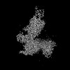 61747MC  9jr2C M: map data used to model this data C: citing same article ( |
|---|---|
| Similar structure data |
-Links
 PDBj
PDBj


- Assembly
| Deposited unit | 
|
|---|---|
| 1 |
|
-Components
-Guanine nucleotide-binding protein ... , 3 types, 3 molecules ABG
| #3: Protein | Mass: 27953.635 Da / Num. of mol.: 1 Source method: isolated from a genetically manipulated source Source: (gene. exp.) Homo sapiens (human) / Production host:   Spodoptera frugiperda (fall armyworm) Spodoptera frugiperda (fall armyworm) |
|---|---|
| #4: Protein | Mass: 37799.270 Da / Num. of mol.: 1 Source method: isolated from a genetically manipulated source Source: (gene. exp.) Spodoptera frugiperda (fall armyworm) / References: UniProt: P54311 |
| #5: Protein | Mass: 7547.685 Da / Num. of mol.: 1 Source method: isolated from a genetically manipulated source Source: (gene. exp.) Spodoptera frugiperda (fall armyworm) / References: UniProt: P63212 |
-Protein / Protein/peptide / Antibody / Sugars / Non-polymers , 5 types, 5 molecules RPS
| #1: Protein | Mass: 63846.227 Da / Num. of mol.: 1 Source method: isolated from a genetically manipulated source Source: (gene. exp.) Homo sapiens (human) / Gene: PTH1R, PTHR, PTHR1 / Cell line (production host): HEK293 / Production host: Homo sapiens (human) / References: UniProt: Q03431 |
|---|---|
| #2: Protein/peptide | Mass: 4123.786 Da / Num. of mol.: 1 / Source method: obtained synthetically / Source: (synth.) Homo sapiens (human) / References: UniProt: P01270 |
| #6: Antibody | Mass: 27720.795 Da / Num. of mol.: 1 Source method: isolated from a genetically manipulated source Source: (gene. exp.) Spodoptera frugiperda (fall armyworm) |
| #7: Polysaccharide | 2-acetamido-2-deoxy-beta-D-glucopyranose-(1-4)-2-acetamido-2-deoxy-beta-D-glucopyranose Source method: isolated from a genetically manipulated source |
| #8: Water | ChemComp-HOH / Mass: 18.015 Da / Num. of mol.: 1 / Source method: isolated from a natural source / Formula: H2O |
-Details
| Has ligand of interest | Y |
|---|---|
| Has protein modification | Y |
-Experimental details
-Experiment
| Experiment | Method: ELECTRON MICROSCOPY |
|---|---|
| EM experiment | Aggregation state: PARTICLE / 3D reconstruction method: single particle reconstruction |
- Sample preparation
| Component |
| ||||||||||||||||||||||||||||||||||||||||||||||||
|---|---|---|---|---|---|---|---|---|---|---|---|---|---|---|---|---|---|---|---|---|---|---|---|---|---|---|---|---|---|---|---|---|---|---|---|---|---|---|---|---|---|---|---|---|---|---|---|---|---|
| Source (natural) |
| ||||||||||||||||||||||||||||||||||||||||||||||||
| Source (recombinant) |
| ||||||||||||||||||||||||||||||||||||||||||||||||
| Buffer solution | pH: 8 | ||||||||||||||||||||||||||||||||||||||||||||||||
| Specimen | Embedding applied: NO / Shadowing applied: NO / Staining applied: NO / Vitrification applied: YES | ||||||||||||||||||||||||||||||||||||||||||||||||
| Vitrification | Cryogen name: ETHANE |
- Electron microscopy imaging
| Experimental equipment |  Model: Titan Krios / Image courtesy: FEI Company |
|---|---|
| Microscopy | Model: TFS KRIOS |
| Electron gun | Electron source:  FIELD EMISSION GUN / Accelerating voltage: 300 kV / Illumination mode: FLOOD BEAM FIELD EMISSION GUN / Accelerating voltage: 300 kV / Illumination mode: FLOOD BEAM |
| Electron lens | Mode: BRIGHT FIELD / Nominal defocus max: 1600 nm / Nominal defocus min: 800 nm |
| Image recording | Electron dose: 50 e/Å2 / Film or detector model: GATAN K3 (6k x 4k) |
- Processing
| EM software | Name: PHENIX / Category: model refinement |
|---|---|
| CTF correction | Type: NONE |
| 3D reconstruction | Resolution: 2.8 Å / Resolution method: FSC 0.143 CUT-OFF / Num. of particles: 259479 / Symmetry type: POINT |
