 Movie
Movie Controller

[English] 日本語
 Yorodumi
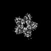
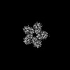
- PDB-9jox: Cryo-EM structure of the light-driven chloride ion-pumping rhodop... -
+ Open data

- Basic information
| Entry | Database: PDB / ID: 9jox | |||||||||||||||||||||||||||
|---|---|---|---|---|---|---|---|---|---|---|---|---|---|---|---|---|---|---|---|---|---|---|---|---|---|---|---|---|
| Title | Cryo-EM structure of the light-driven chloride ion-pumping rhodopsin, NM-R3 | |||||||||||||||||||||||||||
 Components Components | Chloride pumping rhodopsin | |||||||||||||||||||||||||||
 Keywords Keywords | MEMBRANE PROTEIN / chloride ion-pumping rhodopsin RETINAL CELL-FREE SYNTHESIS Bacterial type rhodopsin | |||||||||||||||||||||||||||
| Function / homology |  Function and homology information Function and homology information | |||||||||||||||||||||||||||
| Biological species |  Nonlabens marinus S1-08 (bacteria) Nonlabens marinus S1-08 (bacteria) | |||||||||||||||||||||||||||
| Method | ELECTRON MICROSCOPY / single particle reconstruction / cryo EM / Resolution: 2.5 Å | |||||||||||||||||||||||||||
 Authors Authors | Hosaka, T. / Shirouzu, M. | |||||||||||||||||||||||||||
| Funding support |  Japan, 1items Japan, 1items
| |||||||||||||||||||||||||||
 Citation Citation | Journal: Nat Microbiol / Year: 2025 Title: Carotenoids bind rhodopsins and act as photocycle-accelerating pigments in marine Bacteroidota. Authors: Takayoshi Fujiwara / Toshiaki Hosaka / Masumi Hasegawa-Takano / Yosuke Nishimura / Kento Tominaga / Kaho Mori / Satoshi Nishino / Yuno Takahashi / Tomomi Uchikubo-Kamo / Kazuharu Hanada / ...Authors: Takayoshi Fujiwara / Toshiaki Hosaka / Masumi Hasegawa-Takano / Yosuke Nishimura / Kento Tominaga / Kaho Mori / Satoshi Nishino / Yuno Takahashi / Tomomi Uchikubo-Kamo / Kazuharu Hanada / Takashi Maoka / Shinichi Takaichi / Keiichi Inoue / Mikako Shirouzu / Susumu Yoshizawa / Abstract: Microbial rhodopsins are photoreceptor proteins widely distributed in marine microorganisms that harness light energy and support marine ecosystems. While retinal is typically the sole chromophore in ...Microbial rhodopsins are photoreceptor proteins widely distributed in marine microorganisms that harness light energy and support marine ecosystems. While retinal is typically the sole chromophore in microbial rhodopsins, some proteorhodopsins, which are proton-pumping rhodopsins abundant in the ocean, use carotenoid antennae to transfer light energy to retinal. However, the mechanism by which carotenoids enhance rhodopsin functions remains unclear. Here, using the marine Bacteroidota isolate Nonlabens marinus S1-08, we reconstituted complexes of rhodopsins with the carotenoid myxol and detected energy transfer to retinal in both proteorhodopsin and chloride ion-pumping rhodopsin. Carotenoid binding facilitated light harvesting and accelerated the photocycle, thereby improving the light utilization efficiency of proteorhodopsin. Cryogenic electron microscopy structural analysis further revealed the molecular architecture of the carotenoid-rhodopsin complexes. The ability to bind carotenoids is conserved in rhodopsins of the marine-dominant phylum Bacteroidota, which are widely transcribed in the photic zone. These findings reveal how carotenoids enhance rhodopsin functions in marine Bacteroidota. | |||||||||||||||||||||||||||
| History |
|
- Structure visualization
| Structure viewer | Molecule: MolmilJmol/JSmol |
|---|
- Downloads & links
-Download
| PDBx/mmCIF format | 9jox.cif.gz | 291 KB | Display | PDBx/mmCIF format |
|---|---|---|---|---|
| PDB format | pdb9jox.ent.gz | 231.1 KB | Display | PDB format |
| PDBx/mmJSON format | 9jox.json.gz | Tree view | PDBx/mmJSON format | |
| Others |  Other downloads Other downloads |
-Validation report
| Arichive directory | https://data.pdbj.org/pub/pdb/validation_reports/jo/9joxftp://data.pdbj.org/pub/pdb/validation_reports/jo/9jox | HTTPS FTP |
|---|
-Related structure data
| Related structure data |  61688MC  9jouC  9jovC  9jowC M: map data used to model this data C: citing same article ( |
|---|---|
| Similar structure data |

-Links
 PDBj
PDBj


- Assembly
| Deposited unit | 
|
|---|---|
| 1 |
|
-Components
-Protein , 1 types, 5 molecules ABCDE
| #1: Protein | Mass: 30710.936 Da / Num. of mol.: 5 Source method: isolated from a genetically manipulated source Source: (gene. exp.) Nonlabens marinus S1-08 (bacteria) / Gene: ClR, NMS_1267 / Production host: |
|---|
-Non-polymers , 9 types, 187 molecules 


| #2: Chemical | ChemComp-RET /  Mass: 284.436 Da / Num. of mol.: 5 / Source method: obtained synthetically / Formula: C20H28O / Feature type: SUBJECT OF INVESTIGATION Mass: 284.436 Da / Num. of mol.: 5 / Source method: obtained synthetically / Formula: C20H28O / Feature type: SUBJECT OF INVESTIGATION#3: Chemical | ChemComp-CL /  Mass: 35.453 Da / Num. of mol.: 10 / Source method: obtained synthetically / Formula: Cl / Feature type: SUBJECT OF INVESTIGATION Mass: 35.453 Da / Num. of mol.: 10 / Source method: obtained synthetically / Formula: Cl / Feature type: SUBJECT OF INVESTIGATION#4: Chemical | ChemComp-PC1 /  Mass: 790.145 Da / Num. of mol.: 5 / Source method: obtained synthetically / Formula: C44H88NO8P / Feature type: SUBJECT OF INVESTIGATION / Comment: phospholipid*YM Mass: 790.145 Da / Num. of mol.: 5 / Source method: obtained synthetically / Formula: C44H88NO8P / Feature type: SUBJECT OF INVESTIGATION / Comment: phospholipid*YM#5: Chemical | ChemComp-PLC /  Mass: 622.834 Da / Num. of mol.: 5 / Source method: obtained synthetically / Formula: C32H65NO8P / Feature type: SUBJECT OF INVESTIGATION / Comment: phospholipid*YM Mass: 622.834 Da / Num. of mol.: 5 / Source method: obtained synthetically / Formula: C32H65NO8P / Feature type: SUBJECT OF INVESTIGATION / Comment: phospholipid*YM#6: Chemical |  Mass: 170.335 Da / Num. of mol.: 2 / Source method: obtained synthetically / Formula: C12H26 / Feature type: SUBJECT OF INVESTIGATION Mass: 170.335 Da / Num. of mol.: 2 / Source method: obtained synthetically / Formula: C12H26 / Feature type: SUBJECT OF INVESTIGATION#7: Chemical | ChemComp-R16 /  Mass: 226.441 Da / Num. of mol.: 6 / Source method: obtained synthetically / Formula: C16H34 / Feature type: SUBJECT OF INVESTIGATION Mass: 226.441 Da / Num. of mol.: 6 / Source method: obtained synthetically / Formula: C16H34 / Feature type: SUBJECT OF INVESTIGATION#8: Chemical |  Mass: 254.494 Da / Num. of mol.: 2 / Source method: obtained synthetically / Formula: C18H38 / Feature type: SUBJECT OF INVESTIGATION Mass: 254.494 Da / Num. of mol.: 2 / Source method: obtained synthetically / Formula: C18H38 / Feature type: SUBJECT OF INVESTIGATION#9: Chemical |  Mass: 198.388 Da / Num. of mol.: 2 / Source method: obtained synthetically / Formula: C14H30 / Feature type: SUBJECT OF INVESTIGATION Mass: 198.388 Da / Num. of mol.: 2 / Source method: obtained synthetically / Formula: C14H30 / Feature type: SUBJECT OF INVESTIGATION#10: Water | ChemComp-HOH / | Mass: 18.015 Da / Num. of mol.: 150 / Source method: isolated from a natural source / Formula: H2O |
|---|
-Details
| Has ligand of interest | Y |
|---|---|
| Has protein modification | Y |
-Experimental details
-Experiment
| Experiment | Method: ELECTRON MICROSCOPY |
|---|---|
| EM experiment | Aggregation state: PARTICLE / 3D reconstruction method: single particle reconstruction |
- Sample preparation
| Component | Name: NM-R3 / Type: COMPLEX / Entity ID: #1 / Source: RECOMBINANT |
|---|---|
| Source (natural) | Organism: Nonlabens marinus S1-08 (bacteria) |
| Source (recombinant) | Organism: |
| Buffer solution | pH: 7.2 |
| Specimen | Embedding applied: NO / Shadowing applied: NO / Staining applied: NO / Vitrification applied: YES |
| Vitrification | Cryogen name: ETHANE |
- Electron microscopy imaging
| Experimental equipment |  Model: Titan Krios / Image courtesy: FEI Company |
|---|---|
| Microscopy | Model: FEI TITAN KRIOS |
| Electron gun | Electron source:  FIELD EMISSION GUN / Accelerating voltage: 300 kV / Illumination mode: FLOOD BEAM FIELD EMISSION GUN / Accelerating voltage: 300 kV / Illumination mode: FLOOD BEAM |
| Electron lens | Mode: BRIGHT FIELD / Nominal defocus max: 2000 nm / Nominal defocus min: 800 nm |
| Image recording | Electron dose: 57.534 e/Å2 / Film or detector model: GATAN K3 BIOQUANTUM (6k x 4k) |
- Processing
| CTF correction | Type: PHASE FLIPPING AND AMPLITUDE CORRECTION | ||||||||||||||||||||||||||||||||||||||||||||||||||||||||||||||||||||||||||||||||||||||||||||||||||||||||||
|---|---|---|---|---|---|---|---|---|---|---|---|---|---|---|---|---|---|---|---|---|---|---|---|---|---|---|---|---|---|---|---|---|---|---|---|---|---|---|---|---|---|---|---|---|---|---|---|---|---|---|---|---|---|---|---|---|---|---|---|---|---|---|---|---|---|---|---|---|---|---|---|---|---|---|---|---|---|---|---|---|---|---|---|---|---|---|---|---|---|---|---|---|---|---|---|---|---|---|---|---|---|---|---|---|---|---|---|
| 3D reconstruction | Resolution: 2.5 Å / Resolution method: FSC 0.143 CUT-OFF / Num. of particles: 2081189 / Symmetry type: POINT | ||||||||||||||||||||||||||||||||||||||||||||||||||||||||||||||||||||||||||||||||||||||||||||||||||||||||||
| Refinement | Resolution: 2.5→2.5 Å / Cor.coef. Fo:Fc: 0.85 / SU B: 4.84 / SU ML: 0.096 / ESU R: 0.214 Stereochemistry target values: MAXIMUM LIKELIHOOD WITH PHASES Details: HYDROGENS HAVE BEEN USED IF PRESENT IN THE INPUT
| ||||||||||||||||||||||||||||||||||||||||||||||||||||||||||||||||||||||||||||||||||||||||||||||||||||||||||
| Solvent computation | Solvent model: PARAMETERS FOR MASK CACLULATION | ||||||||||||||||||||||||||||||||||||||||||||||||||||||||||||||||||||||||||||||||||||||||||||||||||||||||||
| Displacement parameters | Biso mean: 117.808 Å2 | ||||||||||||||||||||||||||||||||||||||||||||||||||||||||||||||||||||||||||||||||||||||||||||||||||||||||||
| Refinement step | Cycle: 1 / Total: 11229 | ||||||||||||||||||||||||||||||||||||||||||||||||||||||||||||||||||||||||||||||||||||||||||||||||||||||||||
| Refine LS restraints |
|