 Movie
Movie Controller

[English] 日本語
 Yorodumi
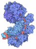
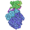
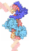
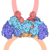
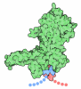
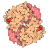
- PDB-9jfd: Human insulin receptor bound with A62-dimer, Pseudo-gamma conformation -
+ Open data

- Basic information
| Entry | Database: PDB / ID: 9jfd | ||||||
|---|---|---|---|---|---|---|---|
| Title | Human insulin receptor bound with A62-dimer, Pseudo-gamma conformation | ||||||
 Components Components |
| ||||||
 Keywords Keywords | MEMBRANE PROTEIN / Signaling / aptamer / agonist / complex | ||||||
| Function / homology |  Function and homology information Function and homology informationregulation of female gonad development / positive regulation of meiotic cell cycle / insulin-like growth factor II binding / positive regulation of developmental growth / insulin receptor complex / insulin-like growth factor I binding / insulin receptor activity / positive regulation of protein-containing complex disassembly / adrenal gland development / dendritic spine maintenance ...regulation of female gonad development / positive regulation of meiotic cell cycle / insulin-like growth factor II binding / positive regulation of developmental growth / insulin receptor complex / insulin-like growth factor I binding / insulin receptor activity / positive regulation of protein-containing complex disassembly / adrenal gland development / dendritic spine maintenance / insulin binding / Signaling by Insulin receptor / PTB domain binding / IRS activation / cargo receptor activity / neuronal cell body membrane / positive regulation of respiratory burst / amyloid-beta clearance / heart morphogenesis / positive regulation of receptor internalization / insulin receptor substrate binding / positive regulation of glycogen biosynthetic process / Signal attenuation / protein kinase activator activity / phosphatidylinositol 3-kinase binding / Insulin receptor recycling / transport across blood-brain barrier / insulin-like growth factor receptor binding / positive regulation of D-glucose import across plasma membrane / positive regulation of mitotic nuclear division / neuron projection maintenance / male gonad development / receptor-mediated endocytosis / positive regulation of glycolytic process / learning / regulation of embryonic development / Insulin receptor signalling cascade / dendrite membrane / memory / receptor protein-tyrosine kinase / caveola / insulin receptor signaling pathway / positive regulation of nitric oxide biosynthetic process / cellular response to insulin stimulus / glucose homeostasis / late endosome / protein autophosphorylation / amyloid-beta binding / PI5P, PP2A and IER3 Regulate PI3K/AKT Signaling / protein tyrosine kinase activity / positive regulation of phosphatidylinositol 3-kinase/protein kinase B signal transduction / positive regulation of canonical NF-kappaB signal transduction / positive regulation of MAPK cascade / signaling receptor complex / lysosome / endosome membrane / positive regulation of cell migration / G protein-coupled receptor signaling pathway / external side of plasma membrane / protein domain specific binding / axon / positive regulation of cell population proliferation / symbiont entry into host cell / regulation of DNA-templated transcription / GTP binding / positive regulation of DNA-templated transcription / protein-containing complex binding / extracellular exosome / ATP binding / membrane / identical protein binding / plasma membrane Similarity search - Function | ||||||
| Biological species |  Homo sapiens (human) Homo sapiens (human) | ||||||
| Method | ELECTRON MICROSCOPY / single particle reconstruction / cryo EM / Resolution: 9.35 Å | ||||||
 Authors Authors | Kim, J. / Na, H. / Yunn, N. / Ryu, S. / Cho, Y. | ||||||
| Funding support |  Korea, Republic Of, 1items Korea, Republic Of, 1items
| ||||||
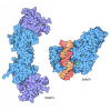
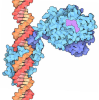
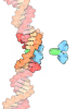
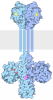
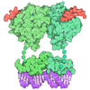
 Citation Citation | Journal: Exp Mol Med / Year: 2025 Title: Structural mechanism of insulin receptor activation by a dimeric aptamer agonist. Authors: Junhong Kim / Hyeonjin Na / Si-Young Choi / Eun Ju Oh / Hyunsook Lee / Sung Ho Ryu / Na-Oh Yunn / Yunje Cho / Abstract: Insulin binding to the insulin receptor (IR) triggers signaling pathways that regulate glucose uptake and cell growth. In previous work, we identified a DNA aptamer, A62, which partially activates ...Insulin binding to the insulin receptor (IR) triggers signaling pathways that regulate glucose uptake and cell growth. In previous work, we identified a DNA aptamer, A62, which partially activates the IR. During engineering aptamers for improved in vivo stability, we discovered that crosslinking two A62 aptamers with linkers of varying lengths led to full phosphorylation of the IR, although activation remained selective to the AKT pathway. Here, to elucidate the mechanism behind this aptamer-induced full activation of the IR, we determined the structure of the IR in complex with a dimeric form of A62 (A62D) linked by an eight-nucleotide connector. We identified three distinct conformations of the IR: arrowhead-shaped, pseudo-arrowhead-shaped and pseudo-gamma-shaped. The pseudo-gamma-shaped conformation closely resembles the structure of a fully active IR bound by a single insulin molecule. In these configurations, only one A62 monomer (A62M) within the A62D dimer binds to the IR dimer. This binding brings the IR monomers into close proximity, promoting intermolecular trans-phosphorylation. Our findings provide valuable structural insights for the development of novel therapeutic strategies targeting the IR. #1: Journal: Exp Mol Med / Year: 2025Title: Structural mechanism of insulin receptor activation by a dimeric aptamer agonist. Authors: Junhong Kim / Hyeonjin Na / Si-Young Choi / Eun Ju Oh / Hyunsook Lee / Sung Ho Ryu / Na-Oh Yunn / Yunje Cho / Abstract: Insulin binding to the insulin receptor (IR) triggers signaling pathways that regulate glucose uptake and cell growth. In previous work, we identified a DNA aptamer, A62, which partially activates ...Insulin binding to the insulin receptor (IR) triggers signaling pathways that regulate glucose uptake and cell growth. In previous work, we identified a DNA aptamer, A62, which partially activates the IR. During engineering aptamers for improved in vivo stability, we discovered that crosslinking two A62 aptamers with linkers of varying lengths led to full phosphorylation of the IR, although activation remained selective to the AKT pathway. Here, to elucidate the mechanism behind this aptamer-induced full activation of the IR, we determined the structure of the IR in complex with a dimeric form of A62 (A62D) linked by an eight-nucleotide connector. We identified three distinct conformations of the IR: arrowhead-shaped, pseudo-arrowhead-shaped and pseudo-gamma-shaped. The pseudo-gamma-shaped conformation closely resembles the structure of a fully active IR bound by a single insulin molecule. In these configurations, only one A62 monomer (A62M) within the A62D dimer binds to the IR dimer. This binding brings the IR monomers into close proximity, promoting intermolecular trans-phosphorylation. Our findings provide valuable structural insights for the development of novel therapeutic strategies targeting the IR. | ||||||
| History |
|
- Structure visualization
| Structure viewer | Molecule: MolmilJmol/JSmol |
|---|
- Downloads & links
-Download
| PDBx/mmCIF format | 9jfd.cif.gz | 305.9 KB | Display | PDBx/mmCIF format |
|---|---|---|---|---|
| PDB format | pdb9jfd.ent.gz | Display | PDB format | |
| PDBx/mmJSON format | 9jfd.json.gz | Tree view | PDBx/mmJSON format | |
| Others |  Other downloads Other downloads |
-Validation report
| Arichive directory | https://data.pdbj.org/pub/pdb/validation_reports/jf/9jfdftp://data.pdbj.org/pub/pdb/validation_reports/jf/9jfd | HTTPS FTP |
|---|
-Related structure data
| Related structure data |  61432MC  9jf9C  9jhsC M: map data used to model this data C: citing same article ( |
|---|---|
| Similar structure data |


-Links
 PDBj
PDBj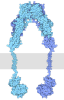












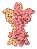



- Assembly
| Deposited unit | 
|
|---|---|
| 1 |
|
-Components
| #1: Protein | Mass: 104848.859 Da / Num. of mol.: 1 Source method: isolated from a genetically manipulated source Source: (gene. exp.) Homo sapiens (human) / Gene: INSR / Production host: Homo sapiens (human)References: UniProt: P06213, receptor protein-tyrosine kinase |
|---|---|
| #2: Protein | Mass: 104812.828 Da / Num. of mol.: 1 Source method: isolated from a genetically manipulated source Source: (gene. exp.) Homo sapiens (human) / Gene: INSR / Production host: Homo sapiens (human)References: UniProt: P06213, receptor protein-tyrosine kinase |
| #3: DNA chain | Mass: 8526.799 Da / Num. of mol.: 1 / Source method: obtained synthetically / Source: (synth.) Homo sapiens (human) |
| Has ligand of interest | N |
| Has protein modification | Y |
-Experimental details
-Experiment
| Experiment | Method: ELECTRON MICROSCOPY |
|---|---|
| EM experiment | Aggregation state: PARTICLE / 3D reconstruction method: single particle reconstruction |
- Sample preparation
| Component | Name: Human insulin receptor with aptamer complex / Type: COMPLEX / Entity ID: all / Source: RECOMBINANT |
|---|---|
| Source (natural) | Organism: Homo sapiens (human) |
| Source (recombinant) | Organism: Homo sapiens (human) |
| Buffer solution | pH: 7.5 |
| Specimen | Embedding applied: NO / Shadowing applied: NO / Staining applied: NO / Vitrification applied: YES |
| Vitrification | Cryogen name: ETHANE |
- Electron microscopy imaging
| Experimental equipment |  Model: Talos Arctica / Image courtesy: FEI Company |
|---|---|
| Microscopy | Model: FEI TALOS ARCTICA |
| Electron gun | Electron source:  FIELD EMISSION GUN / Accelerating voltage: 200 kV / Illumination mode: FLOOD BEAM FIELD EMISSION GUN / Accelerating voltage: 200 kV / Illumination mode: FLOOD BEAM |
| Electron lens | Mode: BRIGHT FIELD / Nominal defocus max: 2000 nm / Nominal defocus min: 1000 nm |
| Image recording | Electron dose: 50 e/Å2 / Film or detector model: GATAN K3 (6k x 4k) |
- Processing
| EM software |
| ||||||||||||
|---|---|---|---|---|---|---|---|---|---|---|---|---|---|
| CTF correction | Type: PHASE FLIPPING ONLY | ||||||||||||
| 3D reconstruction | Resolution: 9.35 Å / Resolution method: FSC 0.143 CUT-OFF / Num. of particles: 37078 / Symmetry type: POINT |
