 Movie
Movie Controller

[English] 日本語
 Yorodumi
- PDB-9jew: Crystal structure of a cupin protein (tm1459, C106V mutant) in ir... -
+ Open data

- Basic information
| Entry | Database: PDB / ID: 9jew | ||||||
|---|---|---|---|---|---|---|---|
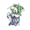
| Title | Crystal structure of a cupin protein (tm1459, C106V mutant) in iron (Fe) substituted form | ||||||
 Components Components | Cupin type-2 domain-containing protein | ||||||
 Keywords Keywords | METAL BINDING PROTEIN / Cupin | ||||||
| Function / homology | Cupin 2, conserved barrel / Cupin domain / RmlC-like cupin domain superfamily / RmlC-like jelly roll fold / metal ion binding / : / Cupin type-2 domain-containing protein Function and homology information Function and homology information | ||||||
| Biological species |   Thermotoga maritima (bacteria) Thermotoga maritima (bacteria) | ||||||
| Method |  X-RAY DIFFRACTION / SYNCHROTRON / MOLECULAR REPLACEMENT / Resolution: 1.08 Å X-RAY DIFFRACTION / SYNCHROTRON / MOLECULAR REPLACEMENT / Resolution: 1.08 Å | ||||||
 Authors Authors | Fujieda, N. / Ichihashi, H. / Kurisu, G. / Itoh, S. | ||||||
| Funding support |  Japan, 1items Japan, 1items
| ||||||
 Citation Citation | Journal: Chem Asian J / Year: 2025 Title: Unusual Self-Hydroxylation in 4-Histidine Tetrad-Supporting Nonheme Iron Center. Authors: Fujieda, N. / Ishihama, K.I. / Ichihashi, H. / Yanagisawa, S. / Kurisu, G. / Itoh, S. | ||||||
| History |
|
- Structure visualization
| Structure viewer | Molecule: MolmilJmol/JSmol |
|---|
- Downloads & links
-Download
| PDBx/mmCIF format | 9jew.cif.gz | 164.4 KB | Display | PDBx/mmCIF format |
|---|---|---|---|---|
| PDB format | pdb9jew.ent.gz | 130.1 KB | Display | PDB format |
| PDBx/mmJSON format | 9jew.json.gz | Tree view | PDBx/mmJSON format | |
| Others |  Other downloads Other downloads |
-Validation report
| Summary document | 9jew_validation.pdf.gz | 1.5 MB | Display | wwPDB validaton report |
|---|---|---|---|---|
| Full document | 9jew_full_validation.pdf.gz | 1.5 MB | Display | |
| Data in XML | 9jew_validation.xml.gz | 16.9 KB | Display | |
| Data in CIF | 9jew_validation.cif.gz | 23.5 KB | Display | |
| Arichive directory | https://data.pdbj.org/pub/pdb/validation_reports/je/9jewftp://data.pdbj.org/pub/pdb/validation_reports/je/9jew | HTTPS FTP |
-Related structure data
| Related structure data |  9jetC  9jeuC  9jevC 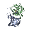 5wsdS S: Starting model for refinement C: citing same article ( |
|---|---|
| Similar structure data |
-Links
 PDBj
PDBj- Assembly
| Deposited unit | 
| ||||||||
|---|---|---|---|---|---|---|---|---|---|
| 1 |
| ||||||||
| Unit cell |
|
-Components
| #1: Protein | Mass: 13423.358 Da / Num. of mol.: 2 / Mutation: C106V Source method: isolated from a genetically manipulated source Source: (gene. exp.) Thermotoga maritima (bacteria) / Gene: TM_1459 / Production host: #2: Chemical |   Mass: 55.845 Da / Num. of mol.: 2 / Source method: obtained synthetically / Formula: Fe / Feature type: SUBJECT OF INVESTIGATION Mass: 55.845 Da / Num. of mol.: 2 / Source method: obtained synthetically / Formula: Fe / Feature type: SUBJECT OF INVESTIGATION#3: Water | ChemComp-HOH / |  Mass: 18.015 Da / Num. of mol.: 291 / Source method: isolated from a natural source / Formula: H2O Mass: 18.015 Da / Num. of mol.: 291 / Source method: isolated from a natural source / Formula: H2OHas ligand of interest | Y | Has protein modification | N | |
|---|
-Experimental details
-Experiment
| Experiment | Method: X-RAY DIFFRACTION / Number of used crystals: 1 |
|---|
- Sample preparation
| Crystal | Density Matthews: 2.07 Å3/Da / Density % sol: 40.69 % |
|---|---|
| Crystal grow | Temperature: 293 K / Method: vapor diffusion, hanging drop / pH: 6 / Details: 25%(w/v) Jeffamine ED-2001, 0.1 M MES |
-Data collection
| Diffraction | Mean temperature: 100 K / Serial crystal experiment: N |
|---|---|
| Diffraction source | Source: SYNCHROTRON / Site: SPring-8 / Beamline: BL44XU / Wavelength: 0.9 Å |
| Detector | Type: DECTRIS EIGER X 16M / Detector: PIXEL / Date: Feb 13, 2023 |
| Radiation | Protocol: SINGLE WAVELENGTH / Monochromatic (M) / Laue (L): M / Scattering type: x-ray |
| Radiation wavelength | Wavelength: 0.9 Å / Relative weight: 1 |
| Reflection | Resolution: 1.08→50 Å / Num. obs: 177500 / % possible obs: 96.6 % / Redundancy: 3.38 % / Rmerge(I) obs: 0.082 / Net I/σ(I): 10.58 |
| Reflection shell | Resolution: 1.08→1.14 Å / Redundancy: 3 % / Rmerge(I) obs: 0.16 / Mean I/σ(I) obs: 4.86 / Num. unique obs: 26186 / % possible all: 88.2 |
- Processing
| Software |
| |||||||||||||||||||||||||||||||||
|---|---|---|---|---|---|---|---|---|---|---|---|---|---|---|---|---|---|---|---|---|---|---|---|---|---|---|---|---|---|---|---|---|---|---|
| Refinement | Method to determine structure: MOLECULAR REPLACEMENT Starting model: 5WSD Resolution: 1.08→30 Å / Cross valid method: FREE R-VALUE / σ(F): 0 / Stereochemistry target values: ENGH AND HUBER
| |||||||||||||||||||||||||||||||||
| Refinement step | Cycle: LAST / Resolution: 1.08→30 Å
| |||||||||||||||||||||||||||||||||
| Refine LS restraints |
| |||||||||||||||||||||||||||||||||
| LS refinement shell | Resolution: 1.08→1.12 Å /
|
