 ムービー
ムービー コントローラー


+ データを開く

- 基本情報
基本情報
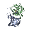
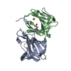
| 登録情報 | データベース: PDB / ID: 9jev | ||||||
|---|---|---|---|---|---|---|---|
| タイトル | Crystal structure of a cupin protein (tm1459) in zinc (Zn) substituted form | ||||||
 要素 要素 | Cupin type-2 domain-containing protein | ||||||
 キーワード キーワード | METAL BINDING PROTEIN / Cupin | ||||||
| 機能・相同性 | Cupin 2, conserved barrel / Cupin domain / RmlC-like cupin domain superfamily / RmlC-like jelly roll fold / metal ion binding / Cupin type-2 domain-containing protein 機能・相同性情報 機能・相同性情報 | ||||||
| 生物種 |   Thermotoga maritima (バクテリア) Thermotoga maritima (バクテリア) | ||||||
| 手法 |  X線回折 / シンクロトロン / 分子置換 / 解像度: 1.12 Å X線回折 / シンクロトロン / 分子置換 / 解像度: 1.12 Å | ||||||
 データ登録者 データ登録者 | Fujieda, N. / Ichihashi, H. / Kurisu, G. / Itoh, S. | ||||||
| 資金援助 |  日本, 1件 日本, 1件
| ||||||
 引用 引用 | ジャーナル: Chem Asian J / 年: 2025 タイトル: Unusual Self-Hydroxylation in 4-Histidine Tetrad-Supporting Nonheme Iron Center. 著者: Fujieda, N. / Ishihama, K.I. / Ichihashi, H. / Yanagisawa, S. / Kurisu, G. / Itoh, S. | ||||||
| 履歴 |
|
- 構造の表示
構造の表示
| 構造ビューア | 分子: MolmilJmol/JSmol |
|---|
- ダウンロードとリンク
-ダウンロード
| PDBx/mmCIF形式 | 9jev.cif.gz | 163.6 KB | 表示 | PDBx/mmCIF形式 |
|---|---|---|---|---|
| PDB形式 | pdb9jev.ent.gz | 129.2 KB | 表示 | PDB形式 |
| PDBx/mmJSON形式 | 9jev.json.gz | ツリー表示 | PDBx/mmJSON形式 | |
| その他 |  その他のダウンロード その他のダウンロード |
-検証レポート
| 文書・要旨 | 9jev_validation.pdf.gz | 1.5 MB | 表示 | wwPDB検証レポート |
|---|---|---|---|---|
| 文書・詳細版 | 9jev_full_validation.pdf.gz | 1.5 MB | 表示 | |
| XML形式データ | 9jev_validation.xml.gz | 17.3 KB | 表示 | |
| CIF形式データ | 9jev_validation.cif.gz | 24.1 KB | 表示 | |
| アーカイブディレクトリ | https://data.pdbj.org/pub/pdb/validation_reports/je/9jevftp://data.pdbj.org/pub/pdb/validation_reports/je/9jev | HTTPS FTP |
-関連構造データ


-リンク
 PDBj
PDBj- 集合体
集合体
| 登録構造単位 | 
| ||||||||
|---|---|---|---|---|---|---|---|---|---|
| 1 |
| ||||||||
| 単位格子 |
|
-要素
| #1: タンパク質 | 分子量: 13459.369 Da / 分子数: 2 / 由来タイプ: 組換発現 由来: (組換発現) Thermotoga maritima (バクテリア)遺伝子: TM_1459 / 発現宿主: #2: 化合物 |   分子量: 65.409 Da / 分子数: 2 / 由来タイプ: 合成 / 式: Zn / タイプ: SUBJECT OF INVESTIGATION 分子量: 65.409 Da / 分子数: 2 / 由来タイプ: 合成 / 式: Zn / タイプ: SUBJECT OF INVESTIGATION#3: 水 | ChemComp-HOH / |  分子量: 18.015 Da / 分子数: 321 / 由来タイプ: 天然 / 式: H2O 分子量: 18.015 Da / 分子数: 321 / 由来タイプ: 天然 / 式: H2O研究の焦点であるリガンドがあるか | Y | Has protein modification | Y | |
|---|
-実験情報
-実験
| 実験 | 手法: X線回折 / 使用した結晶の数: 1 |
|---|
- 試料調製
試料調製
| 結晶 | マシュー密度: 2.08 Å3/Da / 溶媒含有率: 40.97 % |
|---|---|
| 結晶化 | 温度: 293 K / 手法: 蒸気拡散法, ハンギングドロップ法 / pH: 6 / 詳細: 25%(w/v) Jeffamine ED-2001, 0.1 M MES |
-データ収集
| 回折 | 平均測定温度: 100 K / Serial crystal experiment: N |
|---|---|
| 放射光源 | 由来: シンクロトロン / サイト: SPring-8 / ビームライン: BL44XU / 波長: 0.9 Å |
| 検出器 | タイプ: RAYONIX MX300HE / 検出器: CCD / 日付: 2016年5月21日 |
| 放射 | プロトコル: SINGLE WAVELENGTH / 単色(M)・ラウエ(L): M / 散乱光タイプ: x-ray |
| 放射波長 | 波長: 0.9 Å / 相対比: 1 |
| 反射 | 解像度: 1.12→50 Å / Num. obs: 85007 / % possible obs: 99.8 % / 冗長度: 7 % / Rmerge(I) obs: 0.084 / Net I/σ(I): 21.76 |
| 反射 シェル | 解像度: 1.12→1.14 Å / 冗長度: 7 % / Rmerge(I) obs: 0.757 / Mean I/σ(I) obs: 2.64 / Num. unique obs: 4227 / % possible all: 100 |
- 解析
解析
| ソフトウェア |
| |||||||||||||||||||||||||||||||||
|---|---|---|---|---|---|---|---|---|---|---|---|---|---|---|---|---|---|---|---|---|---|---|---|---|---|---|---|---|---|---|---|---|---|---|
| 精密化 | 構造決定の手法: 分子置換 開始モデル: 5WSD 解像度: 1.12→30 Å / 交差検証法: FREE R-VALUE / σ(F): 0 / 立体化学のターゲット値: ENGH AND HUBER
| |||||||||||||||||||||||||||||||||
| 精密化ステップ | サイクル: LAST / 解像度: 1.12→30 Å
| |||||||||||||||||||||||||||||||||
| 拘束条件 |
| |||||||||||||||||||||||||||||||||
| LS精密化 シェル | 解像度: 1.12→1.16 Å /
|
