[English] 日本語
 Yorodumi
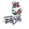
- PDB-9j82: Cryo-EM structure of wild type Aquifex aeolicus RseP in complex w... -
+ Open data
- Basic information
| Entry | Database: PDB / ID: 9j82 | |||||||||||||||||||||||||||
|---|---|---|---|---|---|---|---|---|---|---|---|---|---|---|---|---|---|---|---|---|---|---|---|---|---|---|---|---|
| Title | Cryo-EM structure of wild type Aquifex aeolicus RseP in complex with Fab | |||||||||||||||||||||||||||
 Components Components |
| |||||||||||||||||||||||||||
 Keywords Keywords | HYDROLASE / Protease | |||||||||||||||||||||||||||
| Function / homology |  Function and homology information Function and homology informationHydrolases; Acting on peptide bonds (peptidases); Metalloendopeptidases / metalloendopeptidase activity / proteolysis / metal ion binding / plasma membrane Similarity search - Function | |||||||||||||||||||||||||||
| Biological species |   Aquifex aeolicus VF5 (bacteria) Aquifex aeolicus VF5 (bacteria) | |||||||||||||||||||||||||||
| Method | ELECTRON MICROSCOPY / single particle reconstruction / cryo EM / Resolution: 3.95 Å | |||||||||||||||||||||||||||
 Authors Authors | Asahi, K. / Hirose, M. / Aruga, R. / Kato, T. / Nogi, T. | |||||||||||||||||||||||||||
| Funding support |  Japan, 5items Japan, 5items
| |||||||||||||||||||||||||||
 Citation Citation |  Journal: Sci Adv / Year: 2025 Journal: Sci Adv / Year: 2025Title: Cryo-EM structure of the bacterial intramembrane metalloprotease RseP in the substrate-bound state. Authors: Kikuko Asahi / Mika Hirose / Rie Aruga / Yosuke Shimizu / Michiko Tajiri / Tsubasa Tanaka / Yuriko Adachi / Yukari Tanaka / Mika K Kaneko / Yukinari Kato / Satoko Akashi / Yoshinori Akiyama ...Authors: Kikuko Asahi / Mika Hirose / Rie Aruga / Yosuke Shimizu / Michiko Tajiri / Tsubasa Tanaka / Yuriko Adachi / Yukari Tanaka / Mika K Kaneko / Yukinari Kato / Satoko Akashi / Yoshinori Akiyama / Yohei Hizukuri / Takayuki Kato / Terukazu Nogi /  Abstract: Site-2 proteases (S2Ps), conserved intramembrane metalloproteases that maintain cellular homeostasis, are associated with chronic infection and persistence leading to multidrug resistance in ...Site-2 proteases (S2Ps), conserved intramembrane metalloproteases that maintain cellular homeostasis, are associated with chronic infection and persistence leading to multidrug resistance in bacterial pathogens. A structural model of how S2Ps discriminate and accommodate substrates could help us develop selective antimicrobial agents. We previously proposed that the S2P RseP unwinds helical substrate segments before cleavage, but the mechanism for accommodating a full-length membrane-spanning substrate remained unclear. Our present cryo-EM analysis of RseP (RseP) revealed that a substrate-like membrane protein fragment from the expression host occupied the active site while spanning a transmembrane cavity that is inaccessible via lateral diffusion. Furthermore, in vivo photocrosslinking supported that this substrate accommodation mode is recapitulated on the cell membrane. Our results suggest that the substrate accommodation by threading through a conserved membrane-associated region stabilizes the substrate-complex and contributes to substrate discrimination on the membrane. | |||||||||||||||||||||||||||
| History |
|
- Structure visualization
| Structure viewer | Molecule:  Molmil Molmil Jmol/JSmol Jmol/JSmol |
|---|
- Downloads & links
- Download
| PDBx/mmCIF format |  9j82.cif.gz 9j82.cif.gz | 193.1 KB | Display |  PDBx/mmCIF format PDBx/mmCIF format |
|---|---|---|---|---|
| PDB format |  pdb9j82.ent.gz pdb9j82.ent.gz | 125.9 KB | Display |  PDB format PDB format |
| PDBx/mmJSON format |  9j82.json.gz 9j82.json.gz | Tree view |  PDBx/mmJSON format PDBx/mmJSON format | |
| Others |  Other downloads Other downloads |
-Validation report
| Arichive directory |  https://data.pdbj.org/pub/pdb/validation_reports/j8/9j82 https://data.pdbj.org/pub/pdb/validation_reports/j8/9j82 ftp://data.pdbj.org/pub/pdb/validation_reports/j8/9j82 ftp://data.pdbj.org/pub/pdb/validation_reports/j8/9j82 | HTTPS FTP |
|---|
-Related structure data
| Related structure data |  61213MC  8zayC  9j83C M: map data used to model this data C: citing same article ( |
|---|---|
| Similar structure data | Similarity search - Function & homology  F&H Search F&H Search |
- Links
Links
- Assembly
| Deposited unit | 
|
|---|---|
| 1 |
|
- Components
| #1: Protein | Mass: 49322.203 Da / Num. of mol.: 1 Source method: isolated from a genetically manipulated source Source: (gene. exp.)   Aquifex aeolicus VF5 (bacteria) / Gene: aq_1964 / Production host: Aquifex aeolicus VF5 (bacteria) / Gene: aq_1964 / Production host:  References: UniProt: O67776, Hydrolases; Acting on peptide bonds (peptidases); Metalloendopeptidases |
|---|---|
| #2: Antibody | Mass: 25564.592 Da / Num. of mol.: 1 Source method: isolated from a genetically manipulated source Source: (gene. exp.)   Homo sapiens (human) Homo sapiens (human) |
| #3: Antibody | Mass: 23734.125 Da / Num. of mol.: 1 Source method: isolated from a genetically manipulated source Source: (gene. exp.)   Homo sapiens (human) Homo sapiens (human) |
| #4: Chemical | ChemComp-ZN / |
| Has ligand of interest | N |
| Has protein modification | Y |
-Experimental details
-Experiment
| Experiment | Method: ELECTRON MICROSCOPY |
|---|---|
| EM experiment | Aggregation state: PARTICLE / 3D reconstruction method: single particle reconstruction |
- Sample preparation
| Component |
| ||||||||||||||||||||||||
|---|---|---|---|---|---|---|---|---|---|---|---|---|---|---|---|---|---|---|---|---|---|---|---|---|---|
| Molecular weight |
| ||||||||||||||||||||||||
| Source (natural) |
| ||||||||||||||||||||||||
| Source (recombinant) |
| ||||||||||||||||||||||||
| Buffer solution | pH: 7.4 | ||||||||||||||||||||||||
| Specimen | Embedding applied: NO / Shadowing applied: NO / Staining applied: NO / Vitrification applied: YES | ||||||||||||||||||||||||
| Specimen support | Grid material: GOLD / Grid mesh size: 200 divisions/in. / Grid type: Quantifoil R0.6/1 | ||||||||||||||||||||||||
| Vitrification | Instrument: FEI VITROBOT MARK IV / Cryogen name: ETHANE / Humidity: 95 % / Chamber temperature: 277 K |
- Electron microscopy imaging
| Experimental equipment |  Model: Titan Krios / Image courtesy: FEI Company |
|---|---|
| Microscopy | Model: FEI TITAN KRIOS |
| Electron gun | Electron source:  FIELD EMISSION GUN / Accelerating voltage: 300 kV / Illumination mode: FLOOD BEAM FIELD EMISSION GUN / Accelerating voltage: 300 kV / Illumination mode: FLOOD BEAM |
| Electron lens | Mode: BRIGHT FIELD / Nominal magnification: 105000 X / Calibrated magnification: 103703 X / Nominal defocus max: 1800 nm / Nominal defocus min: 800 nm / Calibrated defocus min: 162 nm / Calibrated defocus max: 3647 nm / Cs: 0.019 mm / C2 aperture diameter: 50 µm / Alignment procedure: COMA FREE |
| Specimen holder | Cryogen: NITROGEN / Specimen holder model: FEI TITAN KRIOS AUTOGRID HOLDER |
| Image recording | Average exposure time: 3.238 sec. / Electron dose: 60 e/Å2 / Film or detector model: GATAN K3 BIOQUANTUM (6k x 4k) / Num. of grids imaged: 1 / Num. of real images: 11400 |
| EM imaging optics | Energyfilter name: GIF Bioquantum / Energyfilter slit width: 20 eV Spherical aberration corrector: CEOS, spherical aberration corrector |
| Image scans | Width: 5760 / Height: 4092 |
- Processing
| EM software |
| ||||||||||||||||||||||||||||||||||||||||||||||||||||||||||||||||||||||
|---|---|---|---|---|---|---|---|---|---|---|---|---|---|---|---|---|---|---|---|---|---|---|---|---|---|---|---|---|---|---|---|---|---|---|---|---|---|---|---|---|---|---|---|---|---|---|---|---|---|---|---|---|---|---|---|---|---|---|---|---|---|---|---|---|---|---|---|---|---|---|---|
| CTF correction | Type: PHASE FLIPPING AND AMPLITUDE CORRECTION | ||||||||||||||||||||||||||||||||||||||||||||||||||||||||||||||||||||||
| Particle selection | Num. of particles selected: 1925238 | ||||||||||||||||||||||||||||||||||||||||||||||||||||||||||||||||||||||
| Symmetry | Point symmetry: C1 (asymmetric) | ||||||||||||||||||||||||||||||||||||||||||||||||||||||||||||||||||||||
| 3D reconstruction | Resolution: 3.95 Å / Resolution method: FSC 0.143 CUT-OFF / Num. of particles: 88124 / Algorithm: FOURIER SPACE / Symmetry type: POINT | ||||||||||||||||||||||||||||||||||||||||||||||||||||||||||||||||||||||
| Atomic model building | Protocol: FLEXIBLE FIT / Space: REAL | ||||||||||||||||||||||||||||||||||||||||||||||||||||||||||||||||||||||
| Atomic model building | 3D fitting-ID: 1
| ||||||||||||||||||||||||||||||||||||||||||||||||||||||||||||||||||||||
| Refinement | Cross valid method: NONE Stereochemistry target values: GeoStd + Monomer Library + CDL v1.2 | ||||||||||||||||||||||||||||||||||||||||||||||||||||||||||||||||||||||
| Displacement parameters | Biso mean: 185.75 Å2 | ||||||||||||||||||||||||||||||||||||||||||||||||||||||||||||||||||||||
| Refine LS restraints |
|
 Movie
Movie Controller


 PDBj
PDBj


