[English] 日本語
Yorodumi
- PDB-8zay: Crystal structure of Fab from mouse monoclonal antibody 4A9 again... -

+
Open data


ID or keywords:

Loading...

-
Basic information

Entry
Database: PDB / ID: 8zay
TitleCrystal structure of Fab from mouse monoclonal antibody 4A9 against Aquifex aeolicus RseP PDZ tandem
Components
  • Light chain of antibody of anti-RseP orthologue from A aeolicus
  • VH-CH1 region of antibody of anti-RseP orthologue from A aeolicus
KeywordsPROTEIN BINDING / Antibody
Biological speciesMus musculus (house mouse)
MethodX-RAY DIFFRACTION / SYNCHROTRON / MOLECULAR REPLACEMENT / Resolution: 2.3 Å
AuthorsAdachi, Y. / Nogi, T.
Funding support Japan, 2items
OrganizationGrant numberCountry
Japan Society for the Promotion of Science (JSPS)23K23825 Japan
Japan Society for the Promotion of Science (JSPS)22H02561 Japan
Citation
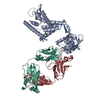
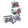
Journal: Sci Adv / Year: 2025
Title: Cryo-EM structure of the bacterial intramembrane metalloprotease RseP in the substrate-bound state.
Authors: Kikuko Asahi / Mika Hirose / Rie Aruga / Yosuke Shimizu / Michiko Tajiri / Tsubasa Tanaka / Yuriko Adachi / Yukari Tanaka / Mika K Kaneko / Yukinari Kato / Satoko Akashi / Yoshinori Akiyama ...Authors: Kikuko Asahi / Mika Hirose / Rie Aruga / Yosuke Shimizu / Michiko Tajiri / Tsubasa Tanaka / Yuriko Adachi / Yukari Tanaka / Mika K Kaneko / Yukinari Kato / Satoko Akashi / Yoshinori Akiyama / Yohei Hizukuri / Takayuki Kato / Terukazu Nogi /
Abstract: Site-2 proteases (S2Ps), conserved intramembrane metalloproteases that maintain cellular homeostasis, are associated with chronic infection and persistence leading to multidrug resistance in ...Site-2 proteases (S2Ps), conserved intramembrane metalloproteases that maintain cellular homeostasis, are associated with chronic infection and persistence leading to multidrug resistance in bacterial pathogens. A structural model of how S2Ps discriminate and accommodate substrates could help us develop selective antimicrobial agents. We previously proposed that the S2P RseP unwinds helical substrate segments before cleavage, but the mechanism for accommodating a full-length membrane-spanning substrate remained unclear. Our present cryo-EM analysis of RseP (RseP) revealed that a substrate-like membrane protein fragment from the expression host occupied the active site while spanning a transmembrane cavity that is inaccessible via lateral diffusion. Furthermore, in vivo photocrosslinking supported that this substrate accommodation mode is recapitulated on the cell membrane. Our results suggest that the substrate accommodation by threading through a conserved membrane-associated region stabilizes the substrate-complex and contributes to substrate discrimination on the membrane.
History
DepositionApr 25, 2024Deposition site: PDBJ / Processing site: PDBJ
Revision 1.0Mar 5, 2025Provider: repository / Type: Initial release
Revision 1.1Mar 12, 2025Group: Database references / Structure summary / Category: citation / citation_author / struct
Item: _citation.journal_volume / _citation.page_first ..._citation.journal_volume / _citation.page_first / _citation.page_last / _citation.pdbx_database_id_PubMed / _citation_author.identifier_ORCID / _struct.title

-
Structure visualization

Structure viewerMolecule:
MolmilJmol/JSmol

Downloads & links

-
Assembly

Deposited unit
H: VH-CH1 region of antibody of anti-RseP orthologue from A aeolicus
I: VH-CH1 region of antibody of anti-RseP orthologue from A aeolicus
L: Light chain of antibody of anti-RseP orthologue from A aeolicus
M: Light chain of antibody of anti-RseP orthologue from A aeolicus
hetero molecules


Theoretical massNumber of molelcules
Total (without water)96,85514
Polymers96,2014
Non-polymers65410
Water4,017223
1
H: VH-CH1 region of antibody of anti-RseP orthologue from A aeolicus
L: Light chain of antibody of anti-RseP orthologue from A aeolicus
hetero molecules


Theoretical massNumber of molelcules
Total (without water)48,4277
Polymers48,1002
Non-polymers3275
Water362
TypeNameSymmetry operationNumber
identity operation1_555x,y,z1
Buried area4260 Å2
ΔGint-165 kcal/mol
Surface area19710 Å2
MethodPISA
2
I: VH-CH1 region of antibody of anti-RseP orthologue from A aeolicus
M: Light chain of antibody of anti-RseP orthologue from A aeolicus
hetero molecules


Theoretical massNumber of molelcules
Total (without water)48,4277
Polymers48,1002
Non-polymers3275
Water362
TypeNameSymmetry operationNumber
identity operation1_555x,y,z1
Buried area4180 Å2
ΔGint-166 kcal/mol
Surface area19700 Å2
MethodPISA
Unit cell
Length a, b, c (Å)51.780, 70.440, 132.460
Angle α, β, γ (deg.)90.000, 96.690, 90.000
Int Tables number4
Space group name H-MP1211
Space group name HallP2yb
Symmetry operation#1: x,y,z
#2: -x,y+1/2,-z

-
Components

#1: Antibody VH-CH1 region of antibody of anti-RseP orthologue from A aeolicus


Mass: 24366.285 Da / Num. of mol.: 2
Source method: isolated from a genetically manipulated source
Source: (gene. exp.) Mus musculus (house mouse) / Production host: Homo sapiens (human)
#2: Antibody Light chain of antibody of anti-RseP orthologue from A aeolicus


Mass: 23734.125 Da / Num. of mol.: 2
Source method: isolated from a genetically manipulated source
Source: (gene. exp.) Mus musculus (house mouse) / Production host: Homo sapiens (human)
#3: Chemical
ChemComp-ZN / ZINC ION


Mass: 65.409 Da / Num. of mol.: 10 / Source method: obtained synthetically / Formula: Zn
#4: Water ChemComp-HOH / water


Mass: 18.015 Da / Num. of mol.: 223 / Source method: isolated from a natural source / Formula: H2O
Has ligand of interestN
Has protein modificationY

-
Experimental details

-
Experiment

ExperimentMethod: X-RAY DIFFRACTION / Number of used crystals: 1

-
Sample preparation

CrystalDensity Matthews: 2.49 Å3/Da / Density % sol: 50.68 %
Crystal growTemperature: 293 K / Method: vapor diffusion, hanging drop
Details: 0.05M Zinc acetate dihydrate, 20% w/v Polyethylene glycol 3350

-
Data collection

DiffractionMean temperature: 100 K / Serial crystal experiment: N
Diffraction sourceSource: SYNCHROTRON / Site: SPring-8 / Beamline: BL32XU / Wavelength: 1 Å
DetectorType: DECTRIS EIGER X 9M / Detector: PIXEL / Date: Jul 24, 2021
RadiationProtocol: SINGLE WAVELENGTH / Monochromatic (M) / Laue (L): M / Scattering type: x-ray
Radiation wavelengthWavelength: 1 Å / Relative weight: 1
ReflectionResolution: 2.3→49.92 Å / Num. obs: 43326 / % possible obs: 100 % / Redundancy: 370.1 % / Biso Wilson estimate: 26.49 Å2 / CC1/2: 0.996 / Rmerge(I) obs: 1.15 / Rpim(I) all: 0.06 / Net I/σ(I): 20.07
Reflection shellResolution: 2.3→2.44 Å / Num. unique obs: 6835 / CC1/2: 0.836

-
Processing

Software
NameVersionClassification
PHENIX1.20.1_4487refinement
XDSdata reduction
XSCALEdata scaling
MOLREPphasing
RefinementMethod to determine structure: MOLECULAR REPLACEMENT / Resolution: 2.3→49.91 Å / SU ML: 0.3666 / Cross valid method: FREE R-VALUE / σ(F): 1.36 / Phase error: 28.2233
Stereochemistry target values: GeoStd + Monomer Library + CDL v1.2
Details: Number of Unique reflections is 82574 when the Bijvoet pairs are separated.
RfactorNum. reflection% reflection
Rfree0.2691 4061 4.92 %
Rwork0.2177 78513 -
obs0.2202 42299 99.96 %
Solvent computationShrinkage radii: 0.9 Å / VDW probe radii: 1.1 Å / Solvent model: FLAT BULK SOLVENT MODEL
Displacement parametersBiso mean: 26.1 Å2
Refinement stepCycle: LAST / Resolution: 2.3→49.91 Å
ProteinNucleic acidLigandSolventTotal
Num. atoms6660 0 10 223 6893
Refine LS restraints
Refine-IDTypeDev idealNumber
X-RAY DIFFRACTIONf_bond_d0.00166822
X-RAY DIFFRACTIONf_angle_d0.44149295
X-RAY DIFFRACTIONf_chiral_restr0.04081046
X-RAY DIFFRACTIONf_plane_restr0.00341187
X-RAY DIFFRACTIONf_dihedral_angle_d5.1863937
LS refinement shell
Resolution (Å)Rfactor RfreeNum. reflection RfreeRfactor RworkNum. reflection RworkRefine-ID% reflection obs (%)
2.3-2.330.44931280.40352700X-RAY DIFFRACTION99.65
2.33-2.360.41561550.36952789X-RAY DIFFRACTION100
2.36-2.390.28871470.30672588X-RAY DIFFRACTION100
2.39-2.420.34561440.2892735X-RAY DIFFRACTION100
2.42-2.450.36131570.28062701X-RAY DIFFRACTION99.97
2.45-2.480.40281200.26972726X-RAY DIFFRACTION100
2.48-2.520.32491380.26162678X-RAY DIFFRACTION100
2.52-2.560.34161490.25272731X-RAY DIFFRACTION100
2.56-2.60.27731240.25982725X-RAY DIFFRACTION100
2.6-2.650.35441520.26182726X-RAY DIFFRACTION100
2.65-2.70.27951140.26592693X-RAY DIFFRACTION100
2.7-2.750.35441480.27692683X-RAY DIFFRACTION100
2.75-2.80.29741320.27872722X-RAY DIFFRACTION100
2.8-2.870.30391340.26172720X-RAY DIFFRACTION100
2.87-2.930.3441480.23222719X-RAY DIFFRACTION100
2.93-3.010.30621380.23082717X-RAY DIFFRACTION100
3.01-3.090.2451410.24242670X-RAY DIFFRACTION100
3.09-3.180.30031540.23112701X-RAY DIFFRACTION100
3.18-3.280.25581440.23222726X-RAY DIFFRACTION100
3.28-3.40.27861340.22272702X-RAY DIFFRACTION100
3.4-3.530.28531730.21442667X-RAY DIFFRACTION100
3.53-3.690.23721500.19182729X-RAY DIFFRACTION100
3.69-3.890.22921320.18812696X-RAY DIFFRACTION100
3.89-4.130.24431740.17122685X-RAY DIFFRACTION100
4.13-4.450.17841280.15522712X-RAY DIFFRACTION100
4.45-4.90.19811120.14052723X-RAY DIFFRACTION100
4.9-5.60.15971370.15622709X-RAY DIFFRACTION100
5.61-7.060.20791310.17912737X-RAY DIFFRACTION100
7.06-49.910.21141230.17552703X-RAY DIFFRACTION99.26

+
About Yorodumi

-
News

-
Feb 9, 2022. New format data for meta-information of EMDB entries

New format data for meta-information of EMDB entries

  • Version 3 of the EMDB header file is now the official format.
  • The previous official version 1.9 will be removed from the archive.

Related info.:EMDB header

External links:wwPDB to switch to version 3 of the EMDB data model

-
Aug 12, 2020. Covid-19 info

Covid-19 info

URL: https://pdbj.org/emnavi/covid19.php

New page: Covid-19 featured information page in EM Navigator.

Related info.:Covid-19 info / Mar 5, 2020. Novel coronavirus structure data

+
Mar 5, 2020. Novel coronavirus structure data

Novel coronavirus structure data

Related info.:Yorodumi Speices / Aug 12, 2020. Covid-19 info

External links:COVID-19 featured content - PDBj / Molecule of the Month (242):Coronavirus Proteases

+
Jan 31, 2019. EMDB accession codes are about to change! (news from PDBe EMDB page)

EMDB accession codes are about to change! (news from PDBe EMDB page)

  • The allocation of 4 digits for EMDB accession codes will soon come to an end. Whilst these codes will remain in use, new EMDB accession codes will include an additional digit and will expand incrementally as the available range of codes is exhausted. The current 4-digit format prefixed with “EMD-” (i.e. EMD-XXXX) will advance to a 5-digit format (i.e. EMD-XXXXX), and so on. It is currently estimated that the 4-digit codes will be depleted around Spring 2019, at which point the 5-digit format will come into force.
  • The EM Navigator/Yorodumi systems omit the EMD- prefix.

Related info.:Q: What is EMD? / ID/Accession-code notation in Yorodumi/EM Navigator

External links:EMDB Accession Codes are Changing Soon! / Contact to PDBj

+
Jul 12, 2017. Major update of PDB

Major update of PDB

  • wwPDB released updated PDB data conforming to the new PDBx/mmCIF dictionary.
  • This is a major update changing the version number from 4 to 5, and with Remediation, in which all the entries are updated.
  • In this update, many items about electron microscopy experimental information are reorganized (e.g. em_software).
  • Now, EM Navigator and Yorodumi are based on the updated data.

External links:wwPDB Remediation / Enriched Model Files Conforming to OneDep Data Standards Now Available in the PDB FTP Archive

-
Yorodumi

Thousand views of thousand structures

  • Yorodumi is a browser for structure data from EMDB, PDB, SASBDB, etc.
  • This page is also the successor to EM Navigator detail page, and also detail information page/front-end page for Omokage search.
  • The word "yorodu" (or yorozu) is an old Japanese word meaning "ten thousand". "mi" (miru) is to see.

Related info.:EMDB / PDB / SASBDB / Comparison of 3 databanks / Yorodumi Search / Aug 31, 2016. New EM Navigator & Yorodumi / Yorodumi Papers / Jmol/JSmol / Function and homology information / Changes in new EM Navigator and Yorodumi

Read more