 Movie
Movie Controller

[English] 日本語
 Yorodumi
- PDB-9j7d: Arabidopsis high-affinity urea transport DUR3 in the inward-facin... -
+ Open data

- Basic information
| Entry | Database: PDB / ID: 9j7d | ||||||
|---|---|---|---|---|---|---|---|
| Title | Arabidopsis high-affinity urea transport DUR3 in the inward-facing open conformation, dimeric state | ||||||
 Components Components | Urea-proton symporter DUR3 | ||||||
 Keywords Keywords | MEMBRANE PROTEIN / DUR3 / high-affinity urea transporter / urea transporter | ||||||
| Function / homology |  Function and homology information Function and homology informationurea binding / urea transmembrane transport / urea transmembrane transporter activity / symporter activity / cellular response to nitrogen starvation / plasma membrane Similarity search - Function | ||||||
| Biological species |  | ||||||
| Method | ELECTRON MICROSCOPY / single particle reconstruction / cryo EM / Resolution: 2.8 Å | ||||||
 Authors Authors | An, W. / Gao, Y. / Zhang, X.C. | ||||||
| Funding support |  China, 1items China, 1items
| ||||||
 Citation Citation | Journal: Nat Commun / Year: 2025 Title: Structural basis of urea transport by Arabidopsis thaliana DUR3. Authors: Weidong An / Yiwei Gao / Laihua Liu / Qinru Bai / Jun Zhao / Yan Zhao / Xuejun C Zhang / Abstract: Urea is a primary nitrogen source used as fertilizer in agricultural plant production and a crucial nitrogen metabolite in plants, playing an essential role in modern agriculture. In plants, DUR3 is ...Urea is a primary nitrogen source used as fertilizer in agricultural plant production and a crucial nitrogen metabolite in plants, playing an essential role in modern agriculture. In plants, DUR3 is a proton-driven high-affinity urea transporter located on the plasma membrane. It not only absorbs external low-concentration urea as a nutrient but also facilitates nitrogen transfer by recovering urea from senescent leaves. Despite its importance, the high-affinity urea transport mechanism in plants remains insufficiently understood. In this study, we determine the structures of Arabidopsis thaliana DUR3 in two different conformations: the inward-facing open state of the apo structure and the occluded urea-bound state, with overall resolutions of 2.8 Å and 3.0 Å, respectively. By comparing these structures and analyzing their functional characteristics, we elucidated how urea molecules are specifically recognized. In the urea-bound structure, we identified key titratable amino acid residues and proposed a model for proton involvement in urea transport based on structural and functional data. This study enhances our understanding of proton-driven urea transport mechanisms in DUR3. | ||||||
| History |
|
- Structure visualization
| Structure viewer | Molecule: MolmilJmol/JSmol |
|---|
- Downloads & links
-Download
| PDBx/mmCIF format | 9j7d.cif.gz | 233.4 KB | Display | PDBx/mmCIF format |
|---|---|---|---|---|
| PDB format | pdb9j7d.ent.gz | 187.4 KB | Display | PDB format |
| PDBx/mmJSON format | 9j7d.json.gz | Tree view | PDBx/mmJSON format | |
| Others |  Other downloads Other downloads |
-Validation report
| Arichive directory | https://data.pdbj.org/pub/pdb/validation_reports/j7/9j7dftp://data.pdbj.org/pub/pdb/validation_reports/j7/9j7d | HTTPS FTP |
|---|
-Related structure data
| Related structure data |  61202MC 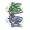 9j7cC M: map data used to model this data C: citing same article ( |
|---|---|
| Similar structure data |

-Links
 PDBj
PDBj- Assembly
| Deposited unit | 
|
|---|---|
| 1 |
|
-Components
| #1: Protein | Mass: 75977.445 Da / Num. of mol.: 2 Source method: isolated from a genetically manipulated source Source: (gene. exp.)  Homo sapiens (human) / References: UniProt: F4KD71 Homo sapiens (human) / References: UniProt: F4KD71#2: Chemical | ChemComp-Y01 /   Mass: 486.726 Da / Num. of mol.: 4 / Source method: isolated from a natural source / Formula: C31H50O4 Mass: 486.726 Da / Num. of mol.: 4 / Source method: isolated from a natural source / Formula: C31H50O4#3: Chemical | ChemComp-C14 / |   Mass: 198.388 Da / Num. of mol.: 1 / Source method: isolated from a natural source / Formula: C14H30 Mass: 198.388 Da / Num. of mol.: 1 / Source method: isolated from a natural source / Formula: C14H30#4: Chemical |   Mass: 226.441 Da / Num. of mol.: 2 / Source method: isolated from a natural source / Formula: C16H34 Mass: 226.441 Da / Num. of mol.: 2 / Source method: isolated from a natural source / Formula: C16H34#5: Water | ChemComp-HOH / |  Mass: 18.015 Da / Num. of mol.: 4 / Source method: isolated from a natural source / Formula: H2O Mass: 18.015 Da / Num. of mol.: 4 / Source method: isolated from a natural source / Formula: H2OHas ligand of interest | N | Has protein modification | Y | |
|---|
-Experimental details
-Experiment
| Experiment | Method: ELECTRON MICROSCOPY |
|---|---|
| EM experiment | Aggregation state: PARTICLE / 3D reconstruction method: single particle reconstruction |
- Sample preparation
| Component | Name: Arabidopsis high-affinity urea transporter / Type: COMPLEX / Entity ID: #1 / Source: RECOMBINANT |
|---|---|
| Source (natural) | Organism: |
| Source (recombinant) | Organism: Homo sapiens (human) |
| Buffer solution | pH: 7.4 |
| Specimen | Conc.: 15 mg/ml / Embedding applied: NO / Shadowing applied: NO / Staining applied: NO / Vitrification applied: YES |
| Vitrification | Cryogen name: ETHANE / Humidity: 100 % / Chamber temperature: 277 K |
- Electron microscopy imaging
| Experimental equipment |  Model: Titan Krios / Image courtesy: FEI Company |
|---|---|
| Microscopy | Model: FEI TITAN KRIOS |
| Electron gun | Electron source:  FIELD EMISSION GUN / Accelerating voltage: 300 kV / Illumination mode: FLOOD BEAM FIELD EMISSION GUN / Accelerating voltage: 300 kV / Illumination mode: FLOOD BEAM |
| Electron lens | Mode: BRIGHT FIELD / Nominal defocus max: 2200 nm / Nominal defocus min: 1200 nm / Cs: 2.7 mm |
| Image recording | Electron dose: 60 e/Å2 / Film or detector model: GATAN K3 (6k x 4k) |
- Processing
| EM software |
| ||||||||||||
|---|---|---|---|---|---|---|---|---|---|---|---|---|---|
| CTF correction | Type: PHASE FLIPPING ONLY | ||||||||||||
| 3D reconstruction | Resolution: 2.8 Å / Resolution method: FSC 0.143 CUT-OFF / Num. of particles: 73561 / Symmetry type: POINT |
