 Movie
Movie Controller

[English] 日本語
 Yorodumi
- PDB-9j4j: Crystal structure of GH9l Inulin fructotransferases(IFTase)incomp... -
+ Open data

- Basic information
| Entry | Database: PDB / ID: 9j4j | ||||||||||||
|---|---|---|---|---|---|---|---|---|---|---|---|---|---|
| Title | Crystal structure of GH9l Inulin fructotransferases(IFTase)incomplex with nystose(F3) | ||||||||||||
 Components Components | DFA-III-forming inulin fructotransferase | ||||||||||||
 Keywords Keywords | CARBOHYDRATE / IFTase / catalytic pathway / HYDROLASE | ||||||||||||
| Function / homology |  Function and homology information Function and homology informationinulin fructotransferase (DFA-III-forming) / inulin fructotransferase (DFA-III-forming) activity / transferase activity Similarity search - Function | ||||||||||||
| Biological species |  Paenarthrobacter aurescens (bacteria) Paenarthrobacter aurescens (bacteria) | ||||||||||||
| Method |  X-RAY DIFFRACTION / MOLECULAR REPLACEMENT / Resolution: 2.803 Å X-RAY DIFFRACTION / MOLECULAR REPLACEMENT / Resolution: 2.803 Å | ||||||||||||
 Authors Authors | Chen, G. / Wang, Z.X. / Yang, Y.Q. / Li, Y.G. / Zhang, T. / Ouyang, S.Y. / Zhang, L. / Chen, Y. / Ruan, X.L. / Miao, M. | ||||||||||||
| Funding support |  China, 3items China, 3items
| ||||||||||||
 Citation Citation | Journal: Int.J.Biol.Macromol. / Year: 2024 Title: Elucidation of the mechanism underlying the sequential catalysis of inulin by fructotransferase. Authors: Chen, G. / Wang, Z.X. / Yang, Y. / Li, Y. / Zhang, T. / Ouyang, S. / Zhang, L. / Chen, Y. / Ruan, X. / Miao, M. | ||||||||||||
| History |
|
- Structure visualization
| Structure viewer | Molecule: MolmilJmol/JSmol |
|---|
- Downloads & links
-Download
| PDBx/mmCIF format | 9j4j.cif.gz | 247.5 KB | Display | PDBx/mmCIF format |
|---|---|---|---|---|
| PDB format | pdb9j4j.ent.gz | 192.4 KB | Display | PDB format |
| PDBx/mmJSON format | 9j4j.json.gz | Tree view | PDBx/mmJSON format | |
| Others |  Other downloads Other downloads |
-Validation report
| Arichive directory | https://data.pdbj.org/pub/pdb/validation_reports/j4/9j4jftp://data.pdbj.org/pub/pdb/validation_reports/j4/9j4j | HTTPS FTP |
|---|
-Related structure data


-Links
 PDBj
PDBj


- Assembly
| Deposited unit | 
| ||||||||
|---|---|---|---|---|---|---|---|---|---|
| 1 |
| ||||||||
| Unit cell |
|
-Components
-Protein / Non-polymers , 2 types, 17 molecules ABC
| #1: Protein | Mass: 43214.438 Da / Num. of mol.: 3 Source method: isolated from a genetically manipulated source Source: (gene. exp.) Paenarthrobacter aurescens (bacteria) / Gene: ift / Production host: References: UniProt: F8QV43, inulin fructotransferase (DFA-III-forming) #6: Water | ChemComp-HOH / | Mass: 18.015 Da / Num. of mol.: 14 / Source method: isolated from a natural source / Formula: H2O |
|---|
-Sugars , 4 types, 8 molecules 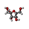
| #2: Polysaccharide | beta-D-fructofuranose-(1-1)-beta-D-fructofuranose Type: oligosaccharide / Mass: 342.297 Da / Num. of mol.: 1 Source method: isolated from a genetically manipulated source | ||||
|---|---|---|---|---|---|
| #3: Polysaccharide | beta-D-fructofuranose-(2-1)-beta-D-fructofuranose-(2-1)-beta-D-fructofuranose-(2-1)-alpha-D-glucopyranose   Source method: isolated from a genetically manipulated source Details: oligosaccharide with reducing-end-to-reducing-end glycosidic bond References: nystose #4: Polysaccharide | beta-D-fructofuranose-(2-1)-beta-D-fructofuranose-(2-1)-beta-D-fructofuranose | Type: oligosaccharide / Mass: 504.438 Da / Num. of mol.: 1 Source method: isolated from a genetically manipulated source #5: Sugar | ChemComp-FRU / |  Type: D-saccharide, beta linking / Mass: 180.156 Da / Num. of mol.: 1 / Source method: obtained synthetically / Formula: C6H12O6 / Feature type: SUBJECT OF INVESTIGATION Type: D-saccharide, beta linking / Mass: 180.156 Da / Num. of mol.: 1 / Source method: obtained synthetically / Formula: C6H12O6 / Feature type: SUBJECT OF INVESTIGATION |
-Details
| Has ligand of interest | Y |
|---|
-Experimental details
-Experiment
| Experiment | Method: X-RAY DIFFRACTION / Number of used crystals: 1 |
|---|
- Sample preparation
| Crystal | Density Matthews: 2.67 Å3/Da / Density meas: 53.91 Mg/m3 / Density % sol: 53.91 % |
|---|---|
| Crystal grow | Temperature: 293 K / Method: vapor diffusion, hanging drop / pH: 8.5 Details: 0.2 M lithium sulfate monohydrate 0.1 M Tris (pH 8.5) 25% (w/v) polyethylene glycol 3350 |
-Data collection
| Diffraction | Mean temperature: 100 K / Serial crystal experiment: N |
|---|---|
| Diffraction source | Source: ROTATING ANODE / Type: RIGAKU FR-E SUPERBRIGHT / Wavelength: 1.54178 Å |
| Detector | Type: RIGAKU SATURN 944+ / Detector: CCD / Date: Dec 31, 2014 |
| Radiation | Protocol: SINGLE WAVELENGTH / Monochromatic (M) / Laue (L): M / Scattering type: x-ray |
| Radiation wavelength | Wavelength: 1.54178 Å / Relative weight: 1 |
| Reflection | Resolution: 2.8→50 Å / Num. obs: 33668 / % possible obs: 100 % / Redundancy: 5.4 % / Rmerge(I) obs: 0.155 / Net I/σ(I): 11.22 |
| Reflection shell | Resolution: 2.8→2.85 Å / Rmerge(I) obs: 0.495 / Num. unique obs: 1682 |
- Processing
| Software |
| ||||||||||||||||||||||||||||||||||||||||||||||||||||||||||||||||||||||||||||||||||||||||||||||||||||||||||||||||||||||||||||||||||||||||||||||||||||||
|---|---|---|---|---|---|---|---|---|---|---|---|---|---|---|---|---|---|---|---|---|---|---|---|---|---|---|---|---|---|---|---|---|---|---|---|---|---|---|---|---|---|---|---|---|---|---|---|---|---|---|---|---|---|---|---|---|---|---|---|---|---|---|---|---|---|---|---|---|---|---|---|---|---|---|---|---|---|---|---|---|---|---|---|---|---|---|---|---|---|---|---|---|---|---|---|---|---|---|---|---|---|---|---|---|---|---|---|---|---|---|---|---|---|---|---|---|---|---|---|---|---|---|---|---|---|---|---|---|---|---|---|---|---|---|---|---|---|---|---|---|---|---|---|---|---|---|---|---|---|---|---|
| Refinement | Method to determine structure: MOLECULAR REPLACEMENT / Resolution: 2.803→48.441 Å / Cor.coef. Fo:Fc: 0.948 / Cor.coef. Fo:Fc free: 0.913 / SU B: 10.101 / SU ML: 0.208 / Cross valid method: FREE R-VALUE / ESU R Free: 0.322 Details: Hydrogens have been added in their riding positions
| ||||||||||||||||||||||||||||||||||||||||||||||||||||||||||||||||||||||||||||||||||||||||||||||||||||||||||||||||||||||||||||||||||||||||||||||||||||||
| Solvent computation | Ion probe radii: 0.8 Å / Shrinkage radii: 0.8 Å / VDW probe radii: 1.2 Å / Solvent model: MASK BULK SOLVENT | ||||||||||||||||||||||||||||||||||||||||||||||||||||||||||||||||||||||||||||||||||||||||||||||||||||||||||||||||||||||||||||||||||||||||||||||||||||||
| Displacement parameters | Biso mean: 31.522 Å2
| ||||||||||||||||||||||||||||||||||||||||||||||||||||||||||||||||||||||||||||||||||||||||||||||||||||||||||||||||||||||||||||||||||||||||||||||||||||||
| Refinement step | Cycle: LAST / Resolution: 2.803→48.441 Å
| ||||||||||||||||||||||||||||||||||||||||||||||||||||||||||||||||||||||||||||||||||||||||||||||||||||||||||||||||||||||||||||||||||||||||||||||||||||||
| Refine LS restraints |
| ||||||||||||||||||||||||||||||||||||||||||||||||||||||||||||||||||||||||||||||||||||||||||||||||||||||||||||||||||||||||||||||||||||||||||||||||||||||
| LS refinement shell |
|
