 Movie
Movie Controller

[English] 日本語
 Yorodumi
- PDB-9j3z: Cryo-EM structure of lysosome cholesterol sencing protein in L state -
+ Open data

- Basic information
| Entry | Database: PDB / ID: 9j3z | |||||||||||||||||||||||||||||||||
|---|---|---|---|---|---|---|---|---|---|---|---|---|---|---|---|---|---|---|---|---|---|---|---|---|---|---|---|---|---|---|---|---|---|---|
| Title | Cryo-EM structure of lysosome cholesterol sencing protein in L state | |||||||||||||||||||||||||||||||||
 Components Components | Lysosomal cholesterol signaling protein,G protein-coupled receptor 155 fusion protein | |||||||||||||||||||||||||||||||||
 Keywords Keywords | LIPID BINDING PROTEIN | |||||||||||||||||||||||||||||||||
| Function / homology |  Function and homology information Function and homology informationprotein localization to lysosome / cellular response to cholesterol / protein-containing complex localization / cholesterol binding / TORC1 signaling / positive regulation of translational initiation / negative regulation of BMP signaling pathway / negative regulation of TORC1 signaling / positive regulation of TORC1 signaling / negative regulation of translational initiation ...protein localization to lysosome / cellular response to cholesterol / protein-containing complex localization / cholesterol binding / TORC1 signaling / positive regulation of translational initiation / negative regulation of BMP signaling pathway / negative regulation of TORC1 signaling / positive regulation of TORC1 signaling / negative regulation of translational initiation / cellular response to amino acid starvation / transmembrane transport / cognition / cytoplasmic translation / intracellular signal transduction / lysosomal membrane / extracellular exosome Similarity search - Function | |||||||||||||||||||||||||||||||||
| Biological species |  Nomascus leucogenys (northern white-cheeked gibbon) Nomascus leucogenys (northern white-cheeked gibbon) Homo sapiens (human) Homo sapiens (human) | |||||||||||||||||||||||||||||||||
| Method | ELECTRON MICROSCOPY / single particle reconstruction / cryo EM / Resolution: 3.5 Å | |||||||||||||||||||||||||||||||||
 Authors Authors | Qian, H.W. / Wang, Z.H. | |||||||||||||||||||||||||||||||||
| Funding support |  China, 1items China, 1items
| |||||||||||||||||||||||||||||||||
 Citation Citation | Journal: Nat Commun / Year: 2025 Title: Structural basis for cholesterol sensing of LYCHOS and its interaction with indoxyl sulfate. Authors: Zhenhua Wang / Jingjing He / Yufan Yang / Yonglin He / Hongwu Qian / Abstract: The lysosome serves as an essential nutrient-sensing hub within the cell, where the mechanistic target of rapamycin complex 1 (mTORC1) is activated. Lysosomal cholesterol signaling (LYCHOS), a ...The lysosome serves as an essential nutrient-sensing hub within the cell, where the mechanistic target of rapamycin complex 1 (mTORC1) is activated. Lysosomal cholesterol signaling (LYCHOS), a lysosome membrane protein, has been identified as a cholesterol sensor that couples cholesterol concentration to mTORC1 activation. However, the molecular basis is unknown. Here, we determine the cryo-electron microscopy (cryo-EM) structure of human LYCHOS at a resolution of 3.1 Å, revealing a cholesterol-like density at the interface between the permease and G-protein coupled receptor (GPCR) domains. Advanced 3D classification reveals two distinct states of LYCHOS. Comparative structural analysis between these two states demonstrated a cholesterol-related movement of GPCR domain relative to permease domain, providing structural insights into how LYCHOS senses lysosomal cholesterol levels. Additionally, we identify indoxyl sulfate (IS) as a binding ligand to the permease domain, confirmed by the LYCHOS-IS complex structure. Overall, our study provides a foundation and indicates additional directions for further investigation of the essential role of LYCHOS in the mTORC1 signaling pathway. | |||||||||||||||||||||||||||||||||
| History |
|
- Structure visualization
| Structure viewer | Molecule: MolmilJmol/JSmol |
|---|
- Downloads & links
-Download
| PDBx/mmCIF format | 9j3z.cif.gz | 212.8 KB | Display | PDBx/mmCIF format |
|---|---|---|---|---|
| PDB format | pdb9j3z.ent.gz | 160.1 KB | Display | PDB format |
| PDBx/mmJSON format | 9j3z.json.gz | Tree view | PDBx/mmJSON format | |
| Others |  Other downloads Other downloads |
-Validation report
| Arichive directory | https://data.pdbj.org/pub/pdb/validation_reports/j3/9j3zftp://data.pdbj.org/pub/pdb/validation_reports/j3/9j3z | HTTPS FTP |
|---|
-Related structure data
| Related structure data |  61128MC 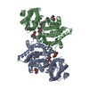 8wr3C  9j3xC 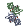 9j40C M: map data used to model this data C: citing same article ( |
|---|---|
| Similar structure data |

-Links
 PDBj
PDBj
- Assembly
| Deposited unit | 
|
|---|---|
| 1 |
|
-Components
| #1: Protein | Mass: 96994.695 Da / Num. of mol.: 2 Source method: isolated from a genetically manipulated source Source: (gene. exp.) Nomascus leucogenys (northern white-cheeked gibbon), (gene. exp.) Homo sapiens (human)Gene: GPR155 / Cell line (production host): HEK293 / Production host: Homo sapiens (human) / References: UniProt: Q7Z3F1, UniProt: G1R1S1#2: Chemical | ChemComp-POV / (   Mass: 760.076 Da / Num. of mol.: 4 / Source method: obtained synthetically / Formula: C42H82NO8P / Feature type: SUBJECT OF INVESTIGATION / Comment: phospholipid*YM Mass: 760.076 Da / Num. of mol.: 4 / Source method: obtained synthetically / Formula: C42H82NO8P / Feature type: SUBJECT OF INVESTIGATION / Comment: phospholipid*YM#3: Sugar |   Type: D-saccharide, beta linking / Mass: 221.208 Da / Num. of mol.: 2 / Source method: obtained synthetically / Formula: C8H15NO6 Type: D-saccharide, beta linking / Mass: 221.208 Da / Num. of mol.: 2 / Source method: obtained synthetically / Formula: C8H15NO6Has ligand of interest | Y | Has protein modification | Y | |
|---|
-Experimental details
-Experiment
| Experiment | Method: ELECTRON MICROSCOPY |
|---|---|
| EM experiment | Aggregation state: PARTICLE / 3D reconstruction method: single particle reconstruction |
- Sample preparation
| Component | Name: Lysosomal cholesterol signaling protein LYCHOS in loose-state Type: COMPLEX / Entity ID: #1 / Source: RECOMBINANT | ||||||||||||
|---|---|---|---|---|---|---|---|---|---|---|---|---|---|
| Molecular weight | Experimental value: NO | ||||||||||||
| Source (natural) |
| ||||||||||||
| Source (recombinant) |
| ||||||||||||
| Buffer solution | pH: 8 | ||||||||||||
| Specimen | Embedding applied: NO / Shadowing applied: NO / Staining applied: NO / Vitrification applied: YES | ||||||||||||
| Vitrification | Cryogen name: ETHANE |
- Electron microscopy imaging
| Experimental equipment |  Model: Titan Krios / Image courtesy: FEI Company |
|---|---|
| Microscopy | Model: FEI TITAN KRIOS |
| Electron gun | Electron source:  FIELD EMISSION GUN / Accelerating voltage: 300 kV / Illumination mode: FLOOD BEAM FIELD EMISSION GUN / Accelerating voltage: 300 kV / Illumination mode: FLOOD BEAM |
| Electron lens | Mode: BRIGHT FIELD / Nominal defocus max: 2000 nm / Nominal defocus min: 1500 nm |
| Image recording | Electron dose: 50 e/Å2 / Film or detector model: GATAN K3 (6k x 4k) |
- Processing
| EM software | Name: PHENIX / Category: model refinement | ||||||||||||||||||||||||
|---|---|---|---|---|---|---|---|---|---|---|---|---|---|---|---|---|---|---|---|---|---|---|---|---|---|
| CTF correction | Type: NONE | ||||||||||||||||||||||||
| 3D reconstruction | Resolution: 3.5 Å / Resolution method: FSC 0.143 CUT-OFF / Num. of particles: 103110 / Symmetry type: POINT | ||||||||||||||||||||||||
| Refine LS restraints |
|
