 Movie
Movie Controller

[English] 日本語
 Yorodumi
- PDB-9ixh: Apg mutant enzyme D448A of the human gut flora K. grimontii TD1 a... -
+ Open data

- Basic information
| Entry | Database: PDB / ID: 9ixh | ||||||
|---|---|---|---|---|---|---|---|
| Title | Apg mutant enzyme D448A of the human gut flora K. grimontii TD1 acarbose hydrolase | ||||||
 Components Components | Maltodextrin glucosidase | ||||||
 Keywords Keywords | HYDROLASE | ||||||
| Function / homology |  Function and homology information Function and homology informationneopullulanase activity / neopullulanase / alpha-1,4-glucosidase activity / alpha-glucosidase / carbohydrate metabolic process / cytoplasm Similarity search - Function | ||||||
| Biological species |  Klebsiella grimontii (bacteria) Klebsiella grimontii (bacteria) | ||||||
| Method |  X-RAY DIFFRACTION / SYNCHROTRON / MOLECULAR REPLACEMENT / Resolution: 2.43 Å X-RAY DIFFRACTION / SYNCHROTRON / MOLECULAR REPLACEMENT / Resolution: 2.43 Å | ||||||
 Authors Authors | Zhou, J.H. / Huang, J.Y. | ||||||
| Funding support | 1items
| ||||||
 Citation Citation | Journal: Nat Commun / Year: 2025 Title: Molecular insights of acarbose metabolization catalyzed by acarbose-preferred glucosidase. Authors: Huang, J. / Shen, Z. / Xiao, X. / Wang, L. / Zhang, J. / Zhou, J. / Gu, Y. | ||||||
| History |
|
- Structure visualization
| Structure viewer | Molecule: MolmilJmol/JSmol |
|---|
- Downloads & links
-Download
| PDBx/mmCIF format | 9ixh.cif.gz | 162.8 KB | Display | PDBx/mmCIF format |
|---|---|---|---|---|
| PDB format | pdb9ixh.ent.gz | 103.3 KB | Display | PDB format |
| PDBx/mmJSON format | 9ixh.json.gz | Tree view | PDBx/mmJSON format | |
| Others |  Other downloads Other downloads |
-Validation report
| Arichive directory | https://data.pdbj.org/pub/pdb/validation_reports/ix/9ixhftp://data.pdbj.org/pub/pdb/validation_reports/ix/9ixh | HTTPS FTP |
|---|
-Related structure data
| Related structure data |  9ivzC  9ixwC  9izeC  9izoC  9unsC  9v0iC  9vd8C  9vdgC  9vdwC 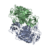 7vt9S C: citing same article ( S: Starting model for refinement |
|---|---|
| Similar structure data |
-Links
 PDBj
PDBj

- Assembly
| Deposited unit | 
| ||||||||||||
|---|---|---|---|---|---|---|---|---|---|---|---|---|---|
| 1 | 
| ||||||||||||
| Unit cell |
| ||||||||||||
| Components on special symmetry positions |
|
-Components
| #1: Protein | Mass: 69064.633 Da / Num. of mol.: 1 / Mutation: D448A Source method: isolated from a genetically manipulated source Source: (gene. exp.) Klebsiella grimontii (bacteria) / Gene: tvaI, malZ, HV064_03130, NCTC9149_05459 / Production host: References: UniProt: A0A7H4P971, neopullulanase, alpha-glucosidase |
|---|---|
| #2: Water | ChemComp-HOH /  Mass: 18.015 Da / Num. of mol.: 160 / Source method: isolated from a natural source / Formula: H2O Mass: 18.015 Da / Num. of mol.: 160 / Source method: isolated from a natural source / Formula: H2O |
| Has protein modification | N |
-Experimental details
-Experiment
| Experiment | Method: X-RAY DIFFRACTION / Number of used crystals: 1 |
|---|
- Sample preparation
| Crystal | Density Matthews: 2.44 Å3/Da / Density % sol: 49.64 % |
|---|---|
| Crystal grow | Temperature: 289.15 K / Method: vapor diffusion, sitting drop / pH: 8.5 Details: 0.7 M Sodium citrate tribasic dihydrate 0.1M Tris, PH=8.5 |
-Data collection
| Diffraction | Mean temperature: 100 K / Serial crystal experiment: N |
|---|---|
| Diffraction source | Source: SYNCHROTRON / Site: SSRF  / Beamline: BL19U1 / Wavelength: 0.98655 Å / Beamline: BL19U1 / Wavelength: 0.98655 Å |
| Detector | Type: DECTRIS PILATUS 6M / Detector: PIXEL / Date: Mar 22, 2024 |
| Radiation | Protocol: SINGLE WAVELENGTH / Monochromatic (M) / Laue (L): M / Scattering type: x-ray |
| Radiation wavelength | Wavelength: 0.98655 Å / Relative weight: 1 |
| Reflection | Resolution: 2.43→20 Å / Num. obs: 27421 / % possible obs: 99.8 % / Redundancy: 8.2 % / Biso Wilson estimate: 45.87 Å2 / CC1/2: 0.998 / Rmerge(I) obs: 0.107 / Net I/σ(I): 13.6 |
| Reflection shell | Resolution: 2.43→2.43 Å / Redundancy: 8.6 % / Rmerge(I) obs: 0.107 / Mean I/σ(I) obs: 2.3 / Num. unique obs: 3026 / CC1/2: 0.791 / % possible all: 100 |
- Processing
| Software |
| |||||||||||||||||||||||||||||||||||||||||||||||||||||||||||||||||||||||||||||
|---|---|---|---|---|---|---|---|---|---|---|---|---|---|---|---|---|---|---|---|---|---|---|---|---|---|---|---|---|---|---|---|---|---|---|---|---|---|---|---|---|---|---|---|---|---|---|---|---|---|---|---|---|---|---|---|---|---|---|---|---|---|---|---|---|---|---|---|---|---|---|---|---|---|---|---|---|---|---|
| Refinement | Method to determine structure: MOLECULAR REPLACEMENT Starting model: 7VT9 Resolution: 2.43→20 Å / SU ML: 0.2882 / Cross valid method: FREE R-VALUE / σ(F): 1.34 / Phase error: 25.5264 Stereochemistry target values: GeoStd + Monomer Library + CDL v1.2
| |||||||||||||||||||||||||||||||||||||||||||||||||||||||||||||||||||||||||||||
| Solvent computation | Shrinkage radii: 0.9 Å / VDW probe radii: 1.11 Å / Solvent model: FLAT BULK SOLVENT MODEL | |||||||||||||||||||||||||||||||||||||||||||||||||||||||||||||||||||||||||||||
| Displacement parameters | Biso mean: 46.04 Å2 | |||||||||||||||||||||||||||||||||||||||||||||||||||||||||||||||||||||||||||||
| Refinement step | Cycle: LAST / Resolution: 2.43→20 Å
| |||||||||||||||||||||||||||||||||||||||||||||||||||||||||||||||||||||||||||||
| Refine LS restraints |
| |||||||||||||||||||||||||||||||||||||||||||||||||||||||||||||||||||||||||||||
| LS refinement shell |
|
