 Movie
Movie Controller

[English] 日本語
 Yorodumi
- PDB-7vt9: CRYSTAL STRUCTURE AT 3.4 ANGSTROMS RESOLUTION OF Maltodextrin glu... -
+ Open data

- Basic information
| Entry | Database: PDB / ID: 7vt9 | |||||||||
|---|---|---|---|---|---|---|---|---|---|---|
| Title | CRYSTAL STRUCTURE AT 3.4 ANGSTROMS RESOLUTION OF Maltodextrin glucosidase, MalZ, FROM Escherichia coli | |||||||||
 Components Components | Maltodextrin glucosidase | |||||||||
 Keywords Keywords | HYDROLASE / Alpha-amylase / Maltodextrin glucosidase | |||||||||
| Function / homology |  Function and homology information Function and homology informationalpha-glucan catabolic process / maltose metabolic process / alpha-1,4-glucosidase activity / alpha-glucosidase / protein homodimerization activity / cytoplasm Similarity search - Function | |||||||||
| Biological species |  | |||||||||
| Method |  X-RAY DIFFRACTION / SYNCHROTRON / MOLECULAR REPLACEMENT / Resolution: 3.3 Å X-RAY DIFFRACTION / SYNCHROTRON / MOLECULAR REPLACEMENT / Resolution: 3.3 Å | |||||||||
 Authors Authors | Ahn, W.-C. / Ahn, Y. / Woo, E.-J. | |||||||||
| Funding support |  Korea, Republic Of, 2items Korea, Republic Of, 2items
| |||||||||
 Citation Citation | Journal: Biochem.Biophys.Res.Commun. / Year: 2022 Title: Dimeric architecture of maltodextrin glucosidase (MalZ) provides insights into the substrate recognition and hydrolysis mechanism. Authors: Ahn, W.C. / An, Y. / Song, K.M. / Park, K.H. / Lee, S.J. / Oh, B.H. / Park, J.T. / Woo, E.J. | |||||||||
| History |
|
- Structure visualization
| Structure viewer | Molecule: MolmilJmol/JSmol |
|---|
- Downloads & links
-Download
| PDBx/mmCIF format | 7vt9.cif.gz | 771.6 KB | Display | PDBx/mmCIF format |
|---|---|---|---|---|
| PDB format | pdb7vt9.ent.gz | 609.8 KB | Display | PDB format |
| PDBx/mmJSON format | 7vt9.json.gz | Tree view | PDBx/mmJSON format | |
| Others |  Other downloads Other downloads |
-Validation report
| Arichive directory | https://data.pdbj.org/pub/pdb/validation_reports/vt/7vt9ftp://data.pdbj.org/pub/pdb/validation_reports/vt/7vt9 | HTTPS FTP |
|---|
-Related structure data
| Related structure data | 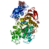 2d0gS S: Starting model for refinement |
|---|---|
| Similar structure data |
-Links
 PDBj
PDBj

- Assembly
| Deposited unit | 
| ||||||||||||||||||
|---|---|---|---|---|---|---|---|---|---|---|---|---|---|---|---|---|---|---|---|
| 1 |
| ||||||||||||||||||
| Unit cell |
| ||||||||||||||||||
| Noncrystallographic symmetry (NCS) | NCS domain:
NCS domain segments: Component-ID: 1 / Ens-ID: ens_1 / Beg auth comp-ID: MET / Beg label comp-ID: MET / End auth comp-ID: ASN / End label comp-ID: ASN / Auth seq-ID: 1 - 604 / Label seq-ID: 1 - 604
NCS oper: (Code: givenMatrix: (0.0246912371563, -0.999667243429, 0.00746627230298), (-0.999665315024, -0.0247474881892, -0.0075378887199), (0.00772015192353, -0.00727765365577, -0.999943715922)Vector: ...NCS oper: (Code: given Matrix: (0.0246912371563, -0.999667243429, 0.00746627230298), Vector: |
-Components
| #1: Protein | Mass: 70406.195 Da / Num. of mol.: 2 Source method: isolated from a genetically manipulated source Source: (gene. exp.) Strain: K12 / Gene: malZ, b0403, JW0393 / Production host: #2: Water | ChemComp-HOH / |  Mass: 18.015 Da / Num. of mol.: 1 / Source method: isolated from a natural source / Formula: H2O Mass: 18.015 Da / Num. of mol.: 1 / Source method: isolated from a natural source / Formula: H2O |
|---|
-Experimental details
-Experiment
| Experiment | Method: X-RAY DIFFRACTION / Number of used crystals: 1 |
|---|
- Sample preparation
| Crystal | Density Matthews: 5.05 Å3/Da / Density % sol: 75.64 % |
|---|---|
| Crystal grow | Temperature: 291 K / Method: vapor diffusion, sitting drop / pH: 7 Details: 20% polyacrylic acid 5100, 100 mM HEPES/Sodium hydroxide pH 7.0 and 20 mM Magnesium chloride |
-Data collection
| Diffraction | Mean temperature: 195 K / Serial crystal experiment: N |
|---|---|
| Diffraction source | Source: SYNCHROTRON / Site: PAL/PLS / Beamline: 7A (6B, 6C1) / Wavelength: 1 Å |
| Detector | Type: ADSC QUANTUM 270 / Detector: CCD / Date: Oct 2, 2013 |
| Radiation | Monochromator: M / Protocol: SINGLE WAVELENGTH / Monochromatic (M) / Laue (L): M / Scattering type: x-ray |
| Radiation wavelength | Wavelength: 1 Å / Relative weight: 1 |
| Reflection | Resolution: 3.3→40.91 Å / Num. obs: 43678 / % possible obs: 99.9 % / Redundancy: 25.4 % / Biso Wilson estimate: 80.22 Å2 / Rmerge(I) obs: 0.355 / Net I/σ(I): 4.02 |
| Reflection shell | Resolution: 3.3→3.418 Å / Redundancy: 14.6 % / Rmerge(I) obs: 0.001 / Num. unique obs: 4169 / % possible all: 99.2 |
- Processing
| Software |
| |||||||||||||||||||||||||||||||||||||||||||||||||||||||||||||||||||||||||||||||||||||||||||||||||||||||||
|---|---|---|---|---|---|---|---|---|---|---|---|---|---|---|---|---|---|---|---|---|---|---|---|---|---|---|---|---|---|---|---|---|---|---|---|---|---|---|---|---|---|---|---|---|---|---|---|---|---|---|---|---|---|---|---|---|---|---|---|---|---|---|---|---|---|---|---|---|---|---|---|---|---|---|---|---|---|---|---|---|---|---|---|---|---|---|---|---|---|---|---|---|---|---|---|---|---|---|---|---|---|---|---|---|---|---|
| Refinement | Method to determine structure: MOLECULAR REPLACEMENT Starting model: 2D0G Resolution: 3.3→40.91 Å / SU ML: 0.6527 / Cross valid method: FREE R-VALUE / σ(F): 1.33 / Phase error: 40.0566 Stereochemistry target values: GeoStd + Monomer Library + CDL v1.2
| |||||||||||||||||||||||||||||||||||||||||||||||||||||||||||||||||||||||||||||||||||||||||||||||||||||||||
| Solvent computation | Shrinkage radii: 0.9 Å / VDW probe radii: 1.11 Å / Solvent model: FLAT BULK SOLVENT MODEL | |||||||||||||||||||||||||||||||||||||||||||||||||||||||||||||||||||||||||||||||||||||||||||||||||||||||||
| Displacement parameters | Biso mean: 120.69 Å2 | |||||||||||||||||||||||||||||||||||||||||||||||||||||||||||||||||||||||||||||||||||||||||||||||||||||||||
| Refinement step | Cycle: LAST / Resolution: 3.3→40.91 Å
| |||||||||||||||||||||||||||||||||||||||||||||||||||||||||||||||||||||||||||||||||||||||||||||||||||||||||
| Refine LS restraints |
| |||||||||||||||||||||||||||||||||||||||||||||||||||||||||||||||||||||||||||||||||||||||||||||||||||||||||
| Refine LS restraints NCS | Type: Torsion NCS / Rms dev position: 1.25651080492 Å | |||||||||||||||||||||||||||||||||||||||||||||||||||||||||||||||||||||||||||||||||||||||||||||||||||||||||
| LS refinement shell |
| |||||||||||||||||||||||||||||||||||||||||||||||||||||||||||||||||||||||||||||||||||||||||||||||||||||||||
| Refinement TLS params. | Method: refined / Origin x: 23.5358538903 Å / Origin y: -23.6310403161 Å / Origin z: -68.7830396425 Å
| |||||||||||||||||||||||||||||||||||||||||||||||||||||||||||||||||||||||||||||||||||||||||||||||||||||||||
| Refinement TLS group | Selection details: all |
