 Movie
Movie Controller


+ Open data

- Basic information
| Entry | Database: PDB / ID: 9isc | ||||||
|---|---|---|---|---|---|---|---|
| Title | Human MTHFD2 in complex with compound 16a | ||||||
 Components Components | Bifunctional methylenetetrahydrofolate dehydrogenase/cyclohydrolase, mitochondrial | ||||||
 Keywords Keywords | OXIDOREDUCTASE / MTHFD2 / methylenetetrahydrofolate dehydrogenase 2 / 1C metabolism / mitochondria / OSIDOREDUCTASE | ||||||
| Function / homology |  Function and homology information Function and homology informationmethylenetetrahydrofolate dehydrogenase (NAD+) / methylenetetrahydrofolate dehydrogenase (NAD+) activity / formate biosynthetic process / methenyltetrahydrofolate cyclohydrolase / methenyltetrahydrofolate cyclohydrolase activity / methylenetetrahydrofolate dehydrogenase (NADP+) activity / Metabolism of folate and pterines / tetrahydrofolate metabolic process / tetrahydrofolate interconversion / phosphate ion binding ...methylenetetrahydrofolate dehydrogenase (NAD+) / methylenetetrahydrofolate dehydrogenase (NAD+) activity / formate biosynthetic process / methenyltetrahydrofolate cyclohydrolase / methenyltetrahydrofolate cyclohydrolase activity / methylenetetrahydrofolate dehydrogenase (NADP+) activity / Metabolism of folate and pterines / tetrahydrofolate metabolic process / tetrahydrofolate interconversion / phosphate ion binding / folic acid metabolic process / mitochondrial matrix / magnesium ion binding / mitochondrion / : Similarity search - Function | ||||||
| Biological species |  Homo sapiens (human) Homo sapiens (human) | ||||||
| Method |  X-RAY DIFFRACTION / SYNCHROTRON / MOLECULAR REPLACEMENT / Resolution: 2.54 Å X-RAY DIFFRACTION / SYNCHROTRON / MOLECULAR REPLACEMENT / Resolution: 2.54 Å | ||||||
 Authors Authors | Lee, L.C. / Wu, S.Y. | ||||||
| Funding support | 1items
| ||||||
 Citation Citation | Journal: J.Med.Chem. / Year: 2024 Title: Development of Potent and Selective Inhibitors of Methylenetetrahydrofolate Dehydrogenase 2 for Targeting Acute Myeloid Leukemia: SAR, Structural Insights, and Biological Characterization. Authors: Chang, H.H. / Lee, L.C. / Hsu, T. / Peng, Y.H. / Huang, C.H. / Yeh, T.K. / Lu, C.T. / Huang, Z.T. / Hsueh, C.C. / Kung, F.C. / Lin, L.M. / Huang, Y.C. / Wang, Y.H. / Li, L.H. / Tang, Y.C. / ...Authors: Chang, H.H. / Lee, L.C. / Hsu, T. / Peng, Y.H. / Huang, C.H. / Yeh, T.K. / Lu, C.T. / Huang, Z.T. / Hsueh, C.C. / Kung, F.C. / Lin, L.M. / Huang, Y.C. / Wang, Y.H. / Li, L.H. / Tang, Y.C. / Chang, L. / Hsieh, C.C. / Jiaang, W.T. / Kuo, C.C. / Wu, S.Y. | ||||||
| History |
|
- Structure visualization
| Structure viewer | Molecule: MolmilJmol/JSmol |
|---|
- Downloads & links
-Download
| PDBx/mmCIF format | 9isc.cif.gz | 129.8 KB | Display | PDBx/mmCIF format |
|---|---|---|---|---|
| PDB format | pdb9isc.ent.gz | Display | PDB format | |
| PDBx/mmJSON format | 9isc.json.gz | Tree view | PDBx/mmJSON format | |
| Others |  Other downloads Other downloads |
-Validation report
| Arichive directory | https://data.pdbj.org/pub/pdb/validation_reports/is/9iscftp://data.pdbj.org/pub/pdb/validation_reports/is/9isc | HTTPS FTP |
|---|
-Related structure data
| Related structure data |  9is9C  9iseC  9islC  9isrC 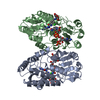 9it3C  9it6C  9itaC 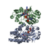 9itdC  9iuoC C: citing same article ( |
|---|---|
| Similar structure data |
-Links
 PDBj
PDBj
- Assembly
| Deposited unit | 
| ||||||||
|---|---|---|---|---|---|---|---|---|---|
| 1 |
| ||||||||
| Unit cell |
|
-Components
| #1: Protein | Mass: 34597.020 Da / Num. of mol.: 2 Source method: isolated from a genetically manipulated source Source: (gene. exp.) Homo sapiens (human) / Gene: MTHFD2, NMDMC / Production host:  References: UniProt: P13995, methylenetetrahydrofolate dehydrogenase (NAD+), methenyltetrahydrofolate cyclohydrolase #2: Chemical |   Mass: 663.425 Da / Num. of mol.: 2 / Source method: obtained synthetically / Formula: C21H27N7O14P2 / Comment: NAD*YM Mass: 663.425 Da / Num. of mol.: 2 / Source method: obtained synthetically / Formula: C21H27N7O14P2 / Comment: NAD*YM#3: Chemical | Mass: 457.403 Da / Num. of mol.: 2 / Source method: obtained synthetically / Formula: C17H19N11O5 / Feature type: SUBJECT OF INVESTIGATION #4: Chemical |   Mass: 94.971 Da / Num. of mol.: 2 / Source method: obtained synthetically / Formula: PO4 Mass: 94.971 Da / Num. of mol.: 2 / Source method: obtained synthetically / Formula: PO4#5: Water | ChemComp-HOH / |  Mass: 18.015 Da / Num. of mol.: 80 / Source method: isolated from a natural source / Formula: H2O Mass: 18.015 Da / Num. of mol.: 80 / Source method: isolated from a natural source / Formula: H2OHas ligand of interest | Y | Has protein modification | N | |
|---|
-Experimental details
-Experiment
| Experiment | Method: X-RAY DIFFRACTION / Number of used crystals: 1 |
|---|
- Sample preparation
| Crystal | Density Matthews: 3.15 Å3/Da / Density % sol: 60.91 % |
|---|---|
| Crystal grow | Temperature: 291.15 K / Method: vapor diffusion, hanging drop / Details: isopropanol, Bis-Tris pH 7.1, PEG 400, PEG 1000 |
-Data collection
| Diffraction | Mean temperature: 100 K / Serial crystal experiment: N | ||||||||||||||||||||||||||||||||||||||||||||||||||||||||||||||||||||||||||||||||||||||||||||||||||||||||||||||
|---|---|---|---|---|---|---|---|---|---|---|---|---|---|---|---|---|---|---|---|---|---|---|---|---|---|---|---|---|---|---|---|---|---|---|---|---|---|---|---|---|---|---|---|---|---|---|---|---|---|---|---|---|---|---|---|---|---|---|---|---|---|---|---|---|---|---|---|---|---|---|---|---|---|---|---|---|---|---|---|---|---|---|---|---|---|---|---|---|---|---|---|---|---|---|---|---|---|---|---|---|---|---|---|---|---|---|---|---|---|---|---|
| Diffraction source | Source: SYNCHROTRON / Site: NSRRC  / Beamline: BL15A1 / Wavelength: 1 Å / Beamline: BL15A1 / Wavelength: 1 Å | ||||||||||||||||||||||||||||||||||||||||||||||||||||||||||||||||||||||||||||||||||||||||||||||||||||||||||||||
| Detector | Type: RAYONIX MX300-HS / Detector: CCD / Date: Mar 21, 2019 | ||||||||||||||||||||||||||||||||||||||||||||||||||||||||||||||||||||||||||||||||||||||||||||||||||||||||||||||
| Radiation | Protocol: SINGLE WAVELENGTH / Monochromatic (M) / Laue (L): M / Scattering type: x-ray | ||||||||||||||||||||||||||||||||||||||||||||||||||||||||||||||||||||||||||||||||||||||||||||||||||||||||||||||
| Radiation wavelength | Wavelength: 1 Å / Relative weight: 1 | ||||||||||||||||||||||||||||||||||||||||||||||||||||||||||||||||||||||||||||||||||||||||||||||||||||||||||||||
| Reflection | Resolution: 2.54→30 Å / Num. obs: 27738 / % possible obs: 98 % / Redundancy: 2.9 % / Rmerge(I) obs: 0.065 / Net I/σ(I): 12.8 | ||||||||||||||||||||||||||||||||||||||||||||||||||||||||||||||||||||||||||||||||||||||||||||||||||||||||||||||
| Reflection shell | Diffraction-ID: 1
|
- Processing
| Software |
| |||||||||||||||||||||||||||||||||||||||||||||||||||||||||||||||||||||||||||||
|---|---|---|---|---|---|---|---|---|---|---|---|---|---|---|---|---|---|---|---|---|---|---|---|---|---|---|---|---|---|---|---|---|---|---|---|---|---|---|---|---|---|---|---|---|---|---|---|---|---|---|---|---|---|---|---|---|---|---|---|---|---|---|---|---|---|---|---|---|---|---|---|---|---|---|---|---|---|---|
| Refinement | Method to determine structure: MOLECULAR REPLACEMENT / Resolution: 2.54→27.18 Å / SU ML: 0.36 / Cross valid method: THROUGHOUT / σ(F): 1.34 / Phase error: 27.2 / Stereochemistry target values: ML
| |||||||||||||||||||||||||||||||||||||||||||||||||||||||||||||||||||||||||||||
| Solvent computation | Shrinkage radii: 0.9 Å / VDW probe radii: 1.11 Å / Solvent model: FLAT BULK SOLVENT MODEL | |||||||||||||||||||||||||||||||||||||||||||||||||||||||||||||||||||||||||||||
| Refinement step | Cycle: LAST / Resolution: 2.54→27.18 Å
| |||||||||||||||||||||||||||||||||||||||||||||||||||||||||||||||||||||||||||||
| Refine LS restraints |
| |||||||||||||||||||||||||||||||||||||||||||||||||||||||||||||||||||||||||||||
| LS refinement shell |
|
