 Movie
Movie Controller

[English] 日本語
 Yorodumi
- PDB-9ijo: X-ray crystal structure of F46C myoglobin with a covalently linke... -
+ Open data

- Basic information
| Entry | Database: PDB / ID: 9ijo | ||||||
|---|---|---|---|---|---|---|---|
| Title | X-ray crystal structure of F46C myoglobin with a covalently linked 4-methyl-2,2'-bipyridine group in complex with Cu2+ | ||||||
 Components Components | Myoglobin | ||||||
 Keywords Keywords | METAL BINDING PROTEIN / heme protein / myoglobin / copper binding | ||||||
| Function / homology |  Function and homology information Function and homology informationOxidoreductases; Acting on other nitrogenous compounds as donors / nitrite reductase activity / sarcoplasm / Oxidoreductases; Acting on a peroxide as acceptor; Peroxidases / removal of superoxide radicals / oxygen carrier activity / peroxidase activity / oxygen binding / heme binding / extracellular exosome / metal ion binding Similarity search - Function | ||||||
| Biological species |  | ||||||
| Method |  X-RAY DIFFRACTION / SYNCHROTRON / MOLECULAR REPLACEMENT / Resolution: 1.53 Å X-RAY DIFFRACTION / SYNCHROTRON / MOLECULAR REPLACEMENT / Resolution: 1.53 Å | ||||||
 Authors Authors | Lin, Y.W. | ||||||
| Funding support |  China, 1items China, 1items
| ||||||
 Citation Citation | Journal: To Be Published Title: X-ray crystal structure of F46C myoglobin with a covalently linked 4-methyl-2,2'-bipyridine group in complex with Cu2+ Authors: Lin, Y.W. | ||||||
| History |
|
- Structure visualization
| Structure viewer | Molecule: MolmilJmol/JSmol |
|---|
- Downloads & links
-Download
| PDBx/mmCIF format | 9ijo.cif.gz | 61.8 KB | Display | PDBx/mmCIF format |
|---|---|---|---|---|
| PDB format | pdb9ijo.ent.gz | Display | PDB format | |
| PDBx/mmJSON format | 9ijo.json.gz | Tree view | PDBx/mmJSON format | |
| Others |  Other downloads Other downloads |
-Validation report
| Summary document | 9ijo_validation.pdf.gz | 1.1 MB | Display | wwPDB validaton report |
|---|---|---|---|---|
| Full document | 9ijo_full_validation.pdf.gz | 1.1 MB | Display | |
| Data in XML | 9ijo_validation.xml.gz | 12.2 KB | Display | |
| Data in CIF | 9ijo_validation.cif.gz | 16.8 KB | Display | |
| Arichive directory | https://data.pdbj.org/pub/pdb/validation_reports/ij/9ijoftp://data.pdbj.org/pub/pdb/validation_reports/ij/9ijo | HTTPS FTP |
-Related structure data
| Related structure data | 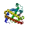 7xc9S S: Starting model for refinement |
|---|---|
| Similar structure data |
-Links
 PDBj
PDBj


- Assembly
| Deposited unit | 
| ||||||||||||
|---|---|---|---|---|---|---|---|---|---|---|---|---|---|
| 1 |
| ||||||||||||
| Unit cell |
|
-Components
| #1: Protein | Mass: 17322.117 Da / Num. of mol.: 1 / Mutation: F46C Source method: isolated from a genetically manipulated source Source: (gene. exp.)  |
|---|---|
| #2: Chemical | ChemComp-HEM /   Mass: 616.487 Da / Num. of mol.: 1 / Source method: obtained synthetically / Formula: C34H32FeN4O4 / Feature type: SUBJECT OF INVESTIGATION Mass: 616.487 Da / Num. of mol.: 1 / Source method: obtained synthetically / Formula: C34H32FeN4O4 / Feature type: SUBJECT OF INVESTIGATION |
| #3: Chemical | ChemComp-A1D9G / Mass: 184.237 Da / Num. of mol.: 1 / Source method: obtained synthetically / Formula: C12H12N2 / Feature type: SUBJECT OF INVESTIGATION |
| #4: Chemical | ChemComp-CU /   Mass: 63.546 Da / Num. of mol.: 1 / Source method: obtained synthetically / Formula: Cu Mass: 63.546 Da / Num. of mol.: 1 / Source method: obtained synthetically / Formula: Cu |
| #5: Water | ChemComp-HOH /  Mass: 18.015 Da / Num. of mol.: 197 / Source method: isolated from a natural source / Formula: H2O Mass: 18.015 Da / Num. of mol.: 197 / Source method: isolated from a natural source / Formula: H2O |
| Has ligand of interest | Y |
| Has protein modification | Y |
-Experimental details
-Experiment
| Experiment | Method: X-RAY DIFFRACTION / Number of used crystals: 1 |
|---|
- Sample preparation
| Crystal | Density Matthews: 2.22 Å3/Da / Density % sol: 44.52 % |
|---|---|
| Crystal grow | Temperature: 289 K / Method: vapor diffusion, hanging drop / Details: sodium acetate, PEG 8000, sodium cacodylate |
-Data collection
| Diffraction | Mean temperature: 100 K / Serial crystal experiment: N |
|---|---|
| Diffraction source | Source: SYNCHROTRON / Site: SSRF / Beamline: BL02U1 / Wavelength: 0.97918 Å |
| Detector | Type: DECTRIS EIGER2 S 9M / Detector: PIXEL / Date: Jun 9, 2024 |
| Radiation | Protocol: SINGLE WAVELENGTH / Monochromatic (M) / Laue (L): M / Scattering type: x-ray |
| Radiation wavelength | Wavelength: 0.97918 Å / Relative weight: 1 |
| Reflection | Resolution: 1.53→30.87 Å / Num. obs: 22848 / % possible obs: 97.07 % / Redundancy: 12.2 % / Biso Wilson estimate: 10.77 Å2 / Rmerge(I) obs: 0.09 / Net I/σ(I): 18.08 |
| Reflection shell | Resolution: 1.53→1.585 Å / Redundancy: 9.7 % / Rmerge(I) obs: 0.49 / Num. unique obs: 2210 / % possible all: 95.41 |
- Processing
| Software |
| |||||||||||||||||||||||||||||||||||||||||||||||||||||||||||||||
|---|---|---|---|---|---|---|---|---|---|---|---|---|---|---|---|---|---|---|---|---|---|---|---|---|---|---|---|---|---|---|---|---|---|---|---|---|---|---|---|---|---|---|---|---|---|---|---|---|---|---|---|---|---|---|---|---|---|---|---|---|---|---|---|---|
| Refinement | Method to determine structure: MOLECULAR REPLACEMENT Starting model: 7XC9 Resolution: 1.53→30.87 Å / SU ML: 0.1473 / Cross valid method: FREE R-VALUE / σ(F): 1.34 / Phase error: 18.1598 Stereochemistry target values: GeoStd + Monomer Library + CDL v1.2
| |||||||||||||||||||||||||||||||||||||||||||||||||||||||||||||||
| Solvent computation | Shrinkage radii: 0.9 Å / VDW probe radii: 1.1 Å / Solvent model: FLAT BULK SOLVENT MODEL | |||||||||||||||||||||||||||||||||||||||||||||||||||||||||||||||
| Displacement parameters | Biso mean: 14.17 Å2 | |||||||||||||||||||||||||||||||||||||||||||||||||||||||||||||||
| Refinement step | Cycle: LAST / Resolution: 1.53→30.87 Å
| |||||||||||||||||||||||||||||||||||||||||||||||||||||||||||||||
| Refine LS restraints |
| |||||||||||||||||||||||||||||||||||||||||||||||||||||||||||||||
| LS refinement shell |
|
