 Movie
Movie Controller


+ Open data

- Basic information
| Entry | Database: PDB / ID: 9hxc | ||||||
|---|---|---|---|---|---|---|---|
| Title | CryoEM structure of Asgard AtubA/B2 microtubule | ||||||
 Components Components |
| ||||||
 Keywords Keywords | STRUCTURAL PROTEIN / Asgard archaea / microtubule / cryoEM / cytomotive filaments / cytoskeleton | ||||||
| Function / homology | GUANOSINE-5'-DIPHOSPHATE Function and homology information Function and homology information | ||||||
| Biological species |  Candidatus Lokiarchaeum ossiferum (archaea) Candidatus Lokiarchaeum ossiferum (archaea) | ||||||
| Method | ELECTRON MICROSCOPY / helical reconstruction / cryo EM / Resolution: 3.3 Å | ||||||
 Authors Authors | Wollweber, F. / Xu, J. / Ponce-Toledo, R.I. / Rodrigues-Oliveira, T. / Malit, J.J.L. / Kokhanovska, A. / Wieczorek, M. / Schleper, C. / Pilhofer, M. | ||||||
| Funding support | European Union, 1items
| ||||||
 Citation Citation | Journal: Cell / Year: 2025 Title: Microtubules in Asgard archaea. Authors: Florian Wollweber / Jingwei Xu / Rafael I Ponce-Toledo / Florina Marxer / Thiago Rodrigues-Oliveira / Anja Pössnecker / Zhen-Hao Luo / Jessie James Limlingan Malit / Anastasiia Kokhanovska ...Authors: Florian Wollweber / Jingwei Xu / Rafael I Ponce-Toledo / Florina Marxer / Thiago Rodrigues-Oliveira / Anja Pössnecker / Zhen-Hao Luo / Jessie James Limlingan Malit / Anastasiia Kokhanovska / Michal Wieczorek / Christa Schleper / Martin Pilhofer /   Abstract: Microtubules are a hallmark of eukaryotes. Archaeal and bacterial homologs of tubulins typically form homopolymers and non-tubular superstructures. The origin of heterodimeric tubulins assembling ...Microtubules are a hallmark of eukaryotes. Archaeal and bacterial homologs of tubulins typically form homopolymers and non-tubular superstructures. The origin of heterodimeric tubulins assembling into microtubules remains unclear. Here, we report the discovery of microtubule-forming tubulins in Asgard archaea, the closest known relatives of eukaryotes. These Asgard tubulins (AtubA/B) are closely related to eukaryotic α/β-tubulins and the enigmatic bacterial tubulins BtubA/B. Proteomics of Candidatus Lokiarchaeum ossiferum showed that AtubA/B were highly expressed. Cryoelectron microscopy structures demonstrate that AtubA/B form eukaryote-like heterodimers, which assembled into 5-protofilament bona fide microtubules in vitro. The additional paralog AtubB2 lacks a nucleotide-binding site and competitively displaced AtubB. These AtubA/B2 heterodimers polymerized into 7-protofilament non-canonical microtubules. In a sub-population of Ca. Lokiarchaeum ossiferum cells, cryo-tomography revealed tubular structures, while expansion microscopy identified AtubA/B cytoskeletal assemblies. Our findings suggest a pre-eukaryotic origin of microtubules and provide a framework for understanding the fundamental principles of microtubule assembly. | ||||||
| History |
|
- Structure visualization
| Structure viewer | Molecule: MolmilJmol/JSmol |
|---|
- Downloads & links
-Download
| PDBx/mmCIF format | 9hxc.cif.gz | 2.2 MB | Display | PDBx/mmCIF format |
|---|---|---|---|---|
| PDB format | pdb9hxc.ent.gz | 1.8 MB | Display | PDB format |
| PDBx/mmJSON format | 9hxc.json.gz | Tree view | PDBx/mmJSON format | |
| Others |  Other downloads Other downloads |
-Validation report
| Arichive directory | https://data.pdbj.org/pub/pdb/validation_reports/hx/9hxcftp://data.pdbj.org/pub/pdb/validation_reports/hx/9hxc | HTTPS FTP |
|---|
-Related structure data
| Related structure data |  52463MC 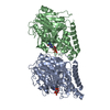 9f6tC  9f6uC 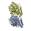 9f6vC M: map data used to model this data C: citing same article ( |
|---|---|
| Similar structure data |


-Links
 PDBj
PDBj
- Assembly
| Deposited unit | 
|
|---|---|
| 1 |
|
-Components
| #1: Protein | Mass: 46706.996 Da / Num. of mol.: 14 Source method: isolated from a genetically manipulated source Source: (gene. exp.) Candidatus Lokiarchaeum ossiferum (archaea)Production host:   Spodoptera frugiperda (fall armyworm) Spodoptera frugiperda (fall armyworm)#2: Protein | Mass: 47882.949 Da / Num. of mol.: 14 Source method: isolated from a genetically manipulated source Source: (gene. exp.) Candidatus Lokiarchaeum ossiferum (archaea)Production host: Spodoptera frugiperda (fall armyworm)#3: Chemical | ChemComp-GDP /   Type: RNA linking / Mass: 443.201 Da / Num. of mol.: 14 / Source method: obtained synthetically / Formula: C10H15N5O11P2 / Feature type: SUBJECT OF INVESTIGATION / Comment: GDP, energy-carrying molecule*YM Type: RNA linking / Mass: 443.201 Da / Num. of mol.: 14 / Source method: obtained synthetically / Formula: C10H15N5O11P2 / Feature type: SUBJECT OF INVESTIGATION / Comment: GDP, energy-carrying molecule*YMHas ligand of interest | Y | Has protein modification | N | |
|---|
-Experimental details
-Experiment
| Experiment | Method: ELECTRON MICROSCOPY |
|---|---|
| EM experiment | Aggregation state: FILAMENT / 3D reconstruction method: helical reconstruction |
- Sample preparation
| Component | Name: microtubule structure of Asgard tubulins AtubA/B2 / Type: COMPLEX / Entity ID: #1-#2 / Source: RECOMBINANT |
|---|---|
| Source (natural) | Organism: Candidatus Lokiarchaeum ossiferum (archaea) |
| Source (recombinant) | Organism: Spodoptera frugiperda (fall armyworm) |
| Buffer solution | pH: 6.8 |
| Specimen | Embedding applied: NO / Shadowing applied: NO / Staining applied: NO / Vitrification applied: YES |
| Vitrification | Cryogen name: ETHANE-PROPANE |
- Electron microscopy imaging
| Experimental equipment |  Model: Titan Krios / Image courtesy: FEI Company |
|---|---|
| Microscopy | Model: TFS KRIOS |
| Electron gun | Electron source:  FIELD EMISSION GUN / Accelerating voltage: 300 kV / Illumination mode: FLOOD BEAM FIELD EMISSION GUN / Accelerating voltage: 300 kV / Illumination mode: FLOOD BEAM |
| Electron lens | Mode: BRIGHT FIELD / Nominal defocus max: 2800 nm / Nominal defocus min: 1200 nm |
| Image recording | Electron dose: 60 e/Å2 / Film or detector model: GATAN K3 (6k x 4k) |
- Processing
| CTF correction | Type: PHASE FLIPPING AND AMPLITUDE CORRECTION |
|---|---|
| Helical symmerty | Angular rotation/subunit: 52.49 ° / Axial rise/subunit: 11.73 Å / Axial symmetry: C1 |
| 3D reconstruction | Resolution: 3.3 Å / Resolution method: FSC 0.143 CUT-OFF / Num. of particles: 39838 / Symmetry type: HELICAL |
