 Movie
Movie Controller

[English] 日本語
 Yorodumi
- PDB-9htd: Peptide-substrate-binding (PSB) domain of human type II collagen ... -
+ Open data

- Basic information
| Entry | Database: PDB / ID: 9htd | |||||||||
|---|---|---|---|---|---|---|---|---|---|---|
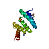
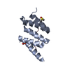
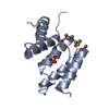
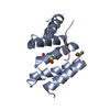
| Title | Peptide-substrate-binding (PSB) domain of human type II collagen prolyl 4-hydroxylase complexed with Pro-Hyp-Gly-Pro-Ala-Gly-Pro-Hyp-Gly. | |||||||||
 Components Components |
| |||||||||
 Keywords Keywords | PROTEIN BINDING / collagen / tetratricopeptide / extracellular matrix | |||||||||
| Function / homology |  Function and homology information Function and homology informationprocollagen-proline 4-dioxygenase / procollagen-proline 4-dioxygenase activity / Collagen biosynthesis and modifying enzymes / L-ascorbic acid binding / electron transfer activity / iron ion binding / endoplasmic reticulum lumen / endoplasmic reticulum / nucleoplasm / cytosol Similarity search - Function | |||||||||
| Biological species |  Homo sapiens (human) Homo sapiens (human) | |||||||||
| Method |  X-RAY DIFFRACTION / SYNCHROTRON / MOLECULAR REPLACEMENT / Resolution: 1.751 Å X-RAY DIFFRACTION / SYNCHROTRON / MOLECULAR REPLACEMENT / Resolution: 1.751 Å | |||||||||
 Authors Authors | Sulu, R. / Rahman, M.M. / Wierenga, R.K. / Koski, M.K. | |||||||||
| Funding support |  Finland, 2items Finland, 2items
| |||||||||
 Citation Citation | Journal: Proteins / Year: 2025 Title: Binding Differences of the Peptide-Substrate-Binding Domain of Collagen Prolyl 4-Hydroxylases I and II for Proline- and Hydroxyproline-Rich Peptides. Authors: Rahman, M.M. / Sulu, R. / Adediran, B. / Tu, H. / Salo, A.M. / Murthy, S. / Myllyharju, J. / Wierenga, R.K. / Koski, M.K. | |||||||||
| History |
|
- Structure visualization
| Structure viewer | Molecule: MolmilJmol/JSmol |
|---|
- Downloads & links
-Download
| PDBx/mmCIF format | 9htd.cif.gz | 56.1 KB | Display | PDBx/mmCIF format |
|---|---|---|---|---|
| PDB format | pdb9htd.ent.gz | 38.8 KB | Display | PDB format |
| PDBx/mmJSON format | 9htd.json.gz | Tree view | PDBx/mmJSON format | |
| Others |  Other downloads Other downloads |
-Validation report
| Arichive directory | https://data.pdbj.org/pub/pdb/validation_reports/ht/9htdftp://data.pdbj.org/pub/pdb/validation_reports/ht/9htd | HTTPS FTP |
|---|
-Related structure data


-Links
 PDBj
PDBj


- Assembly
| Deposited unit | 
| ||||||||
|---|---|---|---|---|---|---|---|---|---|
| 1 |
| ||||||||
| Unit cell |
|
-Components
| #1: Protein | Mass: 11920.266 Da / Num. of mol.: 1 Source method: isolated from a genetically manipulated source Details: N-terminal His-tag. Construct including residues Met142-Glu236. Source: (gene. exp.) Homo sapiens (human) / Gene: P4HA2, UNQ290/PRO330 / Production host:  References: UniProt: O15460, procollagen-proline 4-dioxygenase | ||||||||
|---|---|---|---|---|---|---|---|---|---|
| #2: Protein/peptide | Mass: 777.822 Da / Num. of mol.: 1 / Source method: obtained synthetically Details: The two first N-terminal residues of the peptide are not modelled and not seen in the electron density. Source: (synth.) Homo sapiens (human) | ||||||||
| #3: Chemical | ChemComp-GLY /   Type: peptide linking / Mass: 75.067 Da / Num. of mol.: 4 / Source method: obtained synthetically / Formula: C2H5NO2 Type: peptide linking / Mass: 75.067 Da / Num. of mol.: 4 / Source method: obtained synthetically / Formula: C2H5NO2#4: Chemical | ChemComp-SO4 / |   Mass: 96.063 Da / Num. of mol.: 1 / Source method: obtained synthetically / Formula: SO4 Mass: 96.063 Da / Num. of mol.: 1 / Source method: obtained synthetically / Formula: SO4#5: Water | ChemComp-HOH / |  Mass: 18.015 Da / Num. of mol.: 55 / Source method: isolated from a natural source / Formula: H2O Mass: 18.015 Da / Num. of mol.: 55 / Source method: isolated from a natural source / Formula: H2OHas ligand of interest | N | Has protein modification | Y | |
-Experimental details
-Experiment
| Experiment | Method: X-RAY DIFFRACTION / Number of used crystals: 1 |
|---|
- Sample preparation
| Crystal | Density Matthews: 2.52 Å3/Da / Density % sol: 51.3 % |
|---|---|
| Crystal grow | Temperature: 278 K / Method: vapor diffusion, sitting drop / pH: 5.5 Details: 2M ammonium sulphate, 10% dioxane, 100 mM MES, 2 mM Pro-Hyp-Gly-Pro-Ala-Gly-Pro-Hyp-Gly. Temp details: Cold room temperature is fluctuating between 277 and 279. |
-Data collection
| Diffraction | Mean temperature: 100 K / Serial crystal experiment: N |
|---|---|
| Diffraction source | Source: SYNCHROTRON / Site: PETRA III, EMBL c/o DESY  / Beamline: P14 (MX2) / Wavelength: 0.9763 Å / Beamline: P14 (MX2) / Wavelength: 0.9763 Å |
| Detector | Type: DECTRIS EIGER X 16M / Detector: PIXEL / Date: Sep 9, 2018 / Details: Toroidal mirror |
| Radiation | Monochromator: Si(111) / Protocol: SINGLE WAVELENGTH / Monochromatic (M) / Laue (L): M / Scattering type: x-ray |
| Radiation wavelength | Wavelength: 0.9763 Å / Relative weight: 1 |
| Reflection | Resolution: 1.75→49.06 Å / Num. obs: 12786 / % possible obs: 97.1 % / Redundancy: 6.6 % / Biso Wilson estimate: 27.6 Å2 / CC1/2: 0.998 / Rmerge(I) obs: 0.063 / Rpim(I) all: 0.026 / Net I/σ(I): 14.2 |
| Reflection shell | Resolution: 1.75→1.78 Å / Redundancy: 6.8 % / Rmerge(I) obs: 1.075 / Mean I/σ(I) obs: 1.3 / Num. unique obs: 718 / CC1/2: 0.556 / Rpim(I) all: 0.433 / % possible all: 98.4 |
- Processing
| Software |
| ||||||||||||||||||||||||||||||||||||||||||||||||||||||||||||||||||||||||||||||||||||||||||||||||||||||||||||||||||||||||||||||||||||||||||||||||||||||
|---|---|---|---|---|---|---|---|---|---|---|---|---|---|---|---|---|---|---|---|---|---|---|---|---|---|---|---|---|---|---|---|---|---|---|---|---|---|---|---|---|---|---|---|---|---|---|---|---|---|---|---|---|---|---|---|---|---|---|---|---|---|---|---|---|---|---|---|---|---|---|---|---|---|---|---|---|---|---|---|---|---|---|---|---|---|---|---|---|---|---|---|---|---|---|---|---|---|---|---|---|---|---|---|---|---|---|---|---|---|---|---|---|---|---|---|---|---|---|---|---|---|---|---|---|---|---|---|---|---|---|---|---|---|---|---|---|---|---|---|---|---|---|---|---|---|---|---|---|---|---|---|
| Refinement | Method to determine structure: MOLECULAR REPLACEMENT / Resolution: 1.751→49.06 Å / Cor.coef. Fo:Fc: 0.974 / Cor.coef. Fo:Fc free: 0.963 / SU B: 2.21 / SU ML: 0.067 / Cross valid method: FREE R-VALUE / ESU R: 0.096 / ESU R Free: 0.095 Details: Hydrogens have been added in their riding positions
| ||||||||||||||||||||||||||||||||||||||||||||||||||||||||||||||||||||||||||||||||||||||||||||||||||||||||||||||||||||||||||||||||||||||||||||||||||||||
| Solvent computation | Ion probe radii: 0.8 Å / Shrinkage radii: 0.8 Å / VDW probe radii: 1.2 Å / Solvent model: MASK BULK SOLVENT | ||||||||||||||||||||||||||||||||||||||||||||||||||||||||||||||||||||||||||||||||||||||||||||||||||||||||||||||||||||||||||||||||||||||||||||||||||||||
| Displacement parameters | Biso mean: 39.127 Å2
| ||||||||||||||||||||||||||||||||||||||||||||||||||||||||||||||||||||||||||||||||||||||||||||||||||||||||||||||||||||||||||||||||||||||||||||||||||||||
| Refinement step | Cycle: LAST / Resolution: 1.751→49.06 Å
| ||||||||||||||||||||||||||||||||||||||||||||||||||||||||||||||||||||||||||||||||||||||||||||||||||||||||||||||||||||||||||||||||||||||||||||||||||||||
| Refine LS restraints |
| ||||||||||||||||||||||||||||||||||||||||||||||||||||||||||||||||||||||||||||||||||||||||||||||||||||||||||||||||||||||||||||||||||||||||||||||||||||||
| LS refinement shell |
|
