 Movie
Movie Controller

[English] 日本語
 Yorodumi
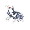
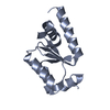
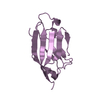
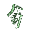
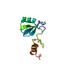
- PDB-6evl: Crystal structure of an unlignaded peptide-substrate-binding doma... -
+ Open data

- Basic information
| Entry | Database: PDB / ID: 6evl | ||||||
|---|---|---|---|---|---|---|---|
| Title | Crystal structure of an unlignaded peptide-substrate-binding domain of human type II collagen prolyl 4-hydroxylase | ||||||
 Components Components | Prolyl 4-hydroxylase subunit alpha-2 | ||||||
 Keywords Keywords | HYDROLASE / tetratricopeptide repeat / collagen synthesis / prolyl 4-hydroxylase | ||||||
| Function / homology |  Function and homology information Function and homology informationprocollagen-proline 4-dioxygenase / procollagen-proline 4-dioxygenase activity / Collagen biosynthesis and modifying enzymes / L-ascorbic acid binding / electron transfer activity / iron ion binding / endoplasmic reticulum lumen / endoplasmic reticulum / nucleoplasm / cytosol Similarity search - Function | ||||||
| Biological species |  Homo sapiens (human) Homo sapiens (human) | ||||||
| Method |  X-RAY DIFFRACTION / MOLECULAR REPLACEMENT / Resolution: 1.87 Å X-RAY DIFFRACTION / MOLECULAR REPLACEMENT / Resolution: 1.87 Å | ||||||
 Authors Authors | Murthy, A.V. / Sulu, R. / Koski, M.K. / Wierenga, R.K. | ||||||
| Funding support |  Finland, 1items Finland, 1items
| ||||||
 Citation Citation | Journal: Protein Sci. / Year: 2018 Title: Structural enzymology binding studies of the peptide-substrate-binding domain of human collagen prolyl 4-hydroxylase (type-II): High affinity peptides have a PxGP sequence motif. Authors: Murthy, A.V. / Sulu, R. / Koski, M.K. / Tu, H. / Anantharajan, J. / Sah-Teli, S.K. / Myllyharju, J. / Wierenga, R.K. | ||||||
| History |
|
- Structure visualization
| Structure viewer | Molecule: MolmilJmol/JSmol |
|---|
- Downloads & links
-Download
| PDBx/mmCIF format | 6evl.cif.gz | 37.6 KB | Display | PDBx/mmCIF format |
|---|---|---|---|---|
| PDB format | pdb6evl.ent.gz | 24.7 KB | Display | PDB format |
| PDBx/mmJSON format | 6evl.json.gz | Tree view | PDBx/mmJSON format | |
| Others |  Other downloads Other downloads |
-Validation report
| Arichive directory | https://data.pdbj.org/pub/pdb/validation_reports/ev/6evlftp://data.pdbj.org/pub/pdb/validation_reports/ev/6evl | HTTPS FTP |
|---|
-Related structure data
| Related structure data | 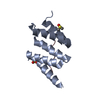 6evmC  6evnC  6evoC 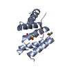 6evpC  2v5fS S: Starting model for refinement C: citing same article ( |
|---|---|
| Similar structure data |


-Links
 PDBj
PDBj


- Assembly
| Deposited unit | 
| ||||||||
|---|---|---|---|---|---|---|---|---|---|
| 1 |
| ||||||||
| Unit cell |
| ||||||||
| Components on special symmetry positions |
|
-Components
| #1: Protein | Mass: 11920.266 Da / Num. of mol.: 1 Source method: isolated from a genetically manipulated source Source: (gene. exp.) Homo sapiens (human) / Gene: P4HA2, UNQ290/PRO330 / Production host:  References: UniProt: O15460, procollagen-proline 4-dioxygenase | ||
|---|---|---|---|
| #2: Chemical | ChemComp-SO4 /   Mass: 96.063 Da / Num. of mol.: 1 / Source method: obtained synthetically / Formula: SO4 Mass: 96.063 Da / Num. of mol.: 1 / Source method: obtained synthetically / Formula: SO4 | ||
| #3: Chemical | ChemComp-DMS /   Mass: 78.133 Da / Num. of mol.: 1 / Source method: obtained synthetically / Formula: C2H6OS / Comment: DMSO, precipitant*YM Mass: 78.133 Da / Num. of mol.: 1 / Source method: obtained synthetically / Formula: C2H6OS / Comment: DMSO, precipitant*YM | ||
| #4: Chemical |   Type: peptide linking / Mass: 75.067 Da / Num. of mol.: 2 / Source method: obtained synthetically / Formula: C2H5NO2 Type: peptide linking / Mass: 75.067 Da / Num. of mol.: 2 / Source method: obtained synthetically / Formula: C2H5NO2#5: Water | ChemComp-HOH / |  Mass: 18.015 Da / Num. of mol.: 122 / Source method: isolated from a natural source / Formula: H2O Mass: 18.015 Da / Num. of mol.: 122 / Source method: isolated from a natural source / Formula: H2O |
-Experimental details
-Experiment
| Experiment | Method: X-RAY DIFFRACTION / Number of used crystals: 1 |
|---|
- Sample preparation
| Crystal | Density Matthews: 2.67 Å3/Da / Density % sol: 54.03 % |
|---|---|
| Crystal grow | Temperature: 277 K / Method: vapor diffusion, sitting drop / pH: 6.5 Details: 2.45 M ammonium sulphate, 10% DMSO, 100 mM MOPS, pH 6.5 |
-Data collection
| Diffraction | Mean temperature: 100 K |
|---|---|
| Diffraction source | Source: ROTATING ANODE / Type: BRUKER AXS MICROSTAR / Wavelength: 1.54 Å |
| Detector | Type: Nonius Kappa CCD / Detector: CCD / Date: Jul 28, 2016 / Details: HELIOS MX MIRRORS |
| Radiation | Protocol: SINGLE WAVELENGTH / Monochromatic (M) / Laue (L): M / Scattering type: x-ray |
| Radiation wavelength | Wavelength: 1.54 Å / Relative weight: 1 |
| Reflection | Resolution: 1.87→48 Å / Num. obs: 10985 / % possible obs: 99.9 % / Redundancy: 14.3 % / Rmerge(I) obs: 0.139 / Rpim(I) all: 0.038 / Net I/σ(I): 15 |
| Reflection shell | Resolution: 1.87→1.9 Å / Redundancy: 5.2 % / Rmerge(I) obs: 0.629 / Mean I/σ(I) obs: 2 / Num. unique obs: 523 / Rpim(I) all: 0.298 / % possible all: 99.8 |
- Processing
| Software |
| ||||||||||||||||||||||||||||||||||||||||||||||||||||||||||||||||||||||||||||||||||||||||||||||||||||||||||||||||
|---|---|---|---|---|---|---|---|---|---|---|---|---|---|---|---|---|---|---|---|---|---|---|---|---|---|---|---|---|---|---|---|---|---|---|---|---|---|---|---|---|---|---|---|---|---|---|---|---|---|---|---|---|---|---|---|---|---|---|---|---|---|---|---|---|---|---|---|---|---|---|---|---|---|---|---|---|---|---|---|---|---|---|---|---|---|---|---|---|---|---|---|---|---|---|---|---|---|---|---|---|---|---|---|---|---|---|---|---|---|---|---|---|---|
| Refinement | Method to determine structure: MOLECULAR REPLACEMENT Starting model: PDB code 2V5F Resolution: 1.87→48 Å / SU ML: 0.22 / Cross valid method: FREE R-VALUE / σ(F): 1.34 / Phase error: 22.95
| ||||||||||||||||||||||||||||||||||||||||||||||||||||||||||||||||||||||||||||||||||||||||||||||||||||||||||||||||
| Solvent computation | Shrinkage radii: 0.9 Å / VDW probe radii: 1.11 Å | ||||||||||||||||||||||||||||||||||||||||||||||||||||||||||||||||||||||||||||||||||||||||||||||||||||||||||||||||
| Refinement step | Cycle: LAST / Resolution: 1.87→48 Å
| ||||||||||||||||||||||||||||||||||||||||||||||||||||||||||||||||||||||||||||||||||||||||||||||||||||||||||||||||
| Refine LS restraints |
| ||||||||||||||||||||||||||||||||||||||||||||||||||||||||||||||||||||||||||||||||||||||||||||||||||||||||||||||||
| LS refinement shell |
|
