 Movie
Movie Controller


+ Open data

- Basic information
| Entry | Database: PDB / ID: 9hpy | ||||||
|---|---|---|---|---|---|---|---|
| Title | Crystal structure of avibactam bound to OXA-57 | ||||||
 Components Components | Beta-lactamase | ||||||
 Keywords Keywords | ANTIMICROBIAL PROTEIN / Class-D Beta-lactamase complex with a inhibitor | ||||||
| Function / homology |  Function and homology information Function and homology informationpenicillin binding / antibiotic catabolic process / beta-lactamase / beta-lactamase activity / response to antibiotic Similarity search - Function | ||||||
| Biological species |  Burkholderia pseudomallei (bacteria) Burkholderia pseudomallei (bacteria) | ||||||
| Method |  X-RAY DIFFRACTION / SYNCHROTRON / MOLECULAR REPLACEMENT / Resolution: 2.48 Å X-RAY DIFFRACTION / SYNCHROTRON / MOLECULAR REPLACEMENT / Resolution: 2.48 Å | ||||||
 Authors Authors | Bragginton, E.C. / Hinchliffe, P. / Spencer, J. | ||||||
| Funding support | 1items
| ||||||
 Citation Citation | Journal: To Be Published Title: Structure and dynamics of Burkholderia pseudomallei OXA-57, a distinctive low efficiency class D beta-lactamase with carbapenem-hydrolyzing activity Authors: Bragginton, E.C. / Colenso, C.K. / Calvopina, K. / Hinchliffe, P. / Shaw, J.M. / Tooke, C.L. / Seng, R. / Chantratita, N. / Schofiled, C.J. / Spencer, J. | ||||||
| History |
|
- Structure visualization
| Structure viewer | Molecule: MolmilJmol/JSmol |
|---|
- Downloads & links
-Download
| PDBx/mmCIF format | 9hpy.cif.gz | 122.3 KB | Display | PDBx/mmCIF format |
|---|---|---|---|---|
| PDB format | pdb9hpy.ent.gz | 83.3 KB | Display | PDB format |
| PDBx/mmJSON format | 9hpy.json.gz | Tree view | PDBx/mmJSON format | |
| Others |  Other downloads Other downloads |
-Validation report
| Arichive directory | https://data.pdbj.org/pub/pdb/validation_reports/hp/9hpyftp://data.pdbj.org/pub/pdb/validation_reports/hp/9hpy | HTTPS FTP |
|---|
-Related structure data


-Links
 PDBj
PDBj


- Assembly
| Deposited unit | 
| ||||||||||||
|---|---|---|---|---|---|---|---|---|---|---|---|---|---|
| 1 |
| ||||||||||||
| Unit cell |
|
-Components
| #1: Protein | Mass: 27033.533 Da / Num. of mol.: 1 Source method: isolated from a genetically manipulated source Source: (gene. exp.) Burkholderia pseudomallei (bacteria) / Gene: oxa-57 / Production host: |
|---|---|
| #2: Chemical | ChemComp-NXL / (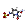  Mass: 267.260 Da / Num. of mol.: 1 / Source method: obtained synthetically / Formula: C7H13N3O6S / Feature type: SUBJECT OF INVESTIGATION / Comment: antibiotic, inhibitor*YM Mass: 267.260 Da / Num. of mol.: 1 / Source method: obtained synthetically / Formula: C7H13N3O6S / Feature type: SUBJECT OF INVESTIGATION / Comment: antibiotic, inhibitor*YM |
| #3: Water | ChemComp-HOH /  Mass: 18.015 Da / Num. of mol.: 9 / Source method: isolated from a natural source / Formula: H2O Mass: 18.015 Da / Num. of mol.: 9 / Source method: isolated from a natural source / Formula: H2O |
| Has ligand of interest | Y |
| Has protein modification | Y |
-Experimental details
-Experiment
| Experiment | Method: X-RAY DIFFRACTION / Number of used crystals: 1 |
|---|
- Sample preparation
| Crystal | Density Matthews: 2.35 Å3/Da / Density % sol: 47.61 % |
|---|---|
| Crystal grow | Temperature: 291 K / Method: vapor diffusion, sitting drop Details: 0.12 M Monosaccharides (0.2 M D-Glucose, 0.2 M D-Mannose, 0.2 M D-Galactose, 0.2 M L-Fucose, 0.2 M D-Xylose, 0.2 M N-Acetyl-D-Glucosamine), 0.1 M Tris; BICINE pH 8.5, 37.5 % (v/v) ...Details: 0.12 M Monosaccharides (0.2 M D-Glucose, 0.2 M D-Mannose, 0.2 M D-Galactose, 0.2 M L-Fucose, 0.2 M D-Xylose, 0.2 M N-Acetyl-D-Glucosamine), 0.1 M Tris; BICINE pH 8.5, 37.5 % (v/v) Precipitant mix (25 % (v/v) MPD, 25 % (w/v) PEG 1000, 35 % (w/v) PEG 3350) Overnight soak in 2mM avibactam. |
-Data collection
| Diffraction | Mean temperature: 100 K / Serial crystal experiment: N |
|---|---|
| Diffraction source | Source: SYNCHROTRON / Site: Diamond  / Beamline: I04-1 / Wavelength: 0.912 Å / Beamline: I04-1 / Wavelength: 0.912 Å |
| Detector | Type: DECTRIS EIGER2 XE 9M / Detector: PIXEL / Date: Feb 3, 2020 |
| Radiation | Protocol: SINGLE WAVELENGTH / Monochromatic (M) / Laue (L): M / Scattering type: x-ray |
| Radiation wavelength | Wavelength: 0.912 Å / Relative weight: 1 |
| Reflection | Resolution: 2.48→55.42 Å / Num. obs: 8900 / % possible obs: 100 % / Redundancy: 12.3 % / CC1/2: 0.999 / Net I/σ(I): 9 |
| Reflection shell | Resolution: 2.48→2.58 Å / Mean I/σ(I) obs: 0.8 / Num. unique obs: 1000 / CC1/2: 0.59 |
- Processing
| Software |
| ||||||||||||||||||||||||||||||||||||||||||||||||||||||||||||||||||||||||||||||||||||||||||||||||||||||||||||||||||||||||||||||||||||||||||||||||||||||
|---|---|---|---|---|---|---|---|---|---|---|---|---|---|---|---|---|---|---|---|---|---|---|---|---|---|---|---|---|---|---|---|---|---|---|---|---|---|---|---|---|---|---|---|---|---|---|---|---|---|---|---|---|---|---|---|---|---|---|---|---|---|---|---|---|---|---|---|---|---|---|---|---|---|---|---|---|---|---|---|---|---|---|---|---|---|---|---|---|---|---|---|---|---|---|---|---|---|---|---|---|---|---|---|---|---|---|---|---|---|---|---|---|---|---|---|---|---|---|---|---|---|---|---|---|---|---|---|---|---|---|---|---|---|---|---|---|---|---|---|---|---|---|---|---|---|---|---|---|---|---|---|
| Refinement | Method to determine structure: MOLECULAR REPLACEMENT / Resolution: 2.48→55.42 Å / SU ML: 0.4531 / Cross valid method: FREE R-VALUE / σ(F): 1.34 / Phase error: 34.2358 Stereochemistry target values: GeoStd + Monomer Library + CDL v1.2
| ||||||||||||||||||||||||||||||||||||||||||||||||||||||||||||||||||||||||||||||||||||||||||||||||||||||||||||||||||||||||||||||||||||||||||||||||||||||
| Solvent computation | Shrinkage radii: 0.9 Å / VDW probe radii: 1.11 Å / Solvent model: FLAT BULK SOLVENT MODEL | ||||||||||||||||||||||||||||||||||||||||||||||||||||||||||||||||||||||||||||||||||||||||||||||||||||||||||||||||||||||||||||||||||||||||||||||||||||||
| Displacement parameters | Biso mean: 76.31 Å2 | ||||||||||||||||||||||||||||||||||||||||||||||||||||||||||||||||||||||||||||||||||||||||||||||||||||||||||||||||||||||||||||||||||||||||||||||||||||||
| Refinement step | Cycle: LAST / Resolution: 2.48→55.42 Å
| ||||||||||||||||||||||||||||||||||||||||||||||||||||||||||||||||||||||||||||||||||||||||||||||||||||||||||||||||||||||||||||||||||||||||||||||||||||||
| Refine LS restraints |
| ||||||||||||||||||||||||||||||||||||||||||||||||||||||||||||||||||||||||||||||||||||||||||||||||||||||||||||||||||||||||||||||||||||||||||||||||||||||
| LS refinement shell |
| ||||||||||||||||||||||||||||||||||||||||||||||||||||||||||||||||||||||||||||||||||||||||||||||||||||||||||||||||||||||||||||||||||||||||||||||||||||||
| Refinement TLS params. | Method: refined / Refine-ID: X-RAY DIFFRACTION
| ||||||||||||||||||||||||||||||||||||||||||||||||||||||||||||||||||||||||||||||||||||||||||||||||||||||||||||||||||||||||||||||||||||||||||||||||||||||
| Refinement TLS group |
|
