 Movie
Movie Controller

[English] 日本語
 Yorodumi
- PDB-9hmf: Periplasmic scaffold of the Campylobacter jejuni flagellar motor ... -
+ Open data

- Basic information
| Entry | Database: PDB / ID: 9hmf | ||||||||||||||||||||||||
|---|---|---|---|---|---|---|---|---|---|---|---|---|---|---|---|---|---|---|---|---|---|---|---|---|---|
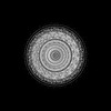
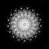
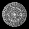
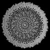
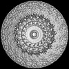
| Title | Periplasmic scaffold of the Campylobacter jejuni flagellar motor (alpha carbon trace) | ||||||||||||||||||||||||
 Components Components |
| ||||||||||||||||||||||||
 Keywords Keywords | STRUCTURAL PROTEIN / molecular machines / flagellar motor / molecular evolution / in situ / scaffold / MOTOR PROTEIN | ||||||||||||||||||||||||
| Function / homology |  Function and homology information Function and homology informationbacterial-type flagellum basal body / bacterial-type flagellum-dependent swarming motility / chemotaxis / membrane / plasma membrane Similarity search - Function | ||||||||||||||||||||||||
| Biological species |   Campylobacter jejuni (Campylobacter) Campylobacter jejuni (Campylobacter) | ||||||||||||||||||||||||
| Method | ELECTRON MICROSCOPY / single particle reconstruction / cryo EM / Resolution: 7.9 Å | ||||||||||||||||||||||||
 Authors Authors | Drobnic, T. / Beeby, M. | ||||||||||||||||||||||||
| Funding support |  United Kingdom, United Kingdom,  France, France,  United States, 7items United States, 7items
| ||||||||||||||||||||||||
 Citation Citation | Journal: Nat Microbiol / Year: 2025 Title: In situ structure of a bacterial flagellar motor at subnanometre resolution reveals adaptations for increased torque. Authors: Tina Drobnič / Eli J Cohen / Thomas Calcraft / Mona Alzheimer / Kathrin Froschauer / Sarah Svensson / William H Hoffmann / Nanki Singh / Sriram G Garg / Louie D Henderson / Trishant R ...Authors: Tina Drobnič / Eli J Cohen / Thomas Calcraft / Mona Alzheimer / Kathrin Froschauer / Sarah Svensson / William H Hoffmann / Nanki Singh / Sriram G Garg / Louie D Henderson / Trishant R Umrekar / Andrea Nans / Deborah Ribardo / Francesco Pedaci / Ashley L Nord / Georg K A Hochberg / David R Hendrixson / Cynthia M Sharma / Peter B Rosenthal / Morgan Beeby /   Abstract: The bacterial flagellar motor, which spins a helical propeller for propulsion, has undergone evolutionary diversification across bacterial species, often involving the addition of structures ...The bacterial flagellar motor, which spins a helical propeller for propulsion, has undergone evolutionary diversification across bacterial species, often involving the addition of structures associated with increasing torque for motility in viscous environments. Understanding how such structures function and have evolved is hampered by challenges in visualizing motors in situ. Here we developed a Campylobacter jejuni minicell system for in situ cryogenic electron microscopy imaging and single-particle analysis of its motor, one of the most complex flagellar motors known, to subnanometre resolution. Focusing on the large periplasmic structures which are essential for increasing torque, our structural data, interpreted with molecular models, show that the basal disk comprises concentric rings of FlgP. The medial disk is a lattice of PflC with PflD, while the proximal disk is a rim of PflB attached to spokes of PflA. PflAB dimerization is essential for proximal disk assembly, recruiting FliL to scaffold more stator complexes at a wider radius which increases torque. We also acquired insights into universal principles of flagellar torque generation. This in situ approach is broadly applicable to other membrane-residing bacterial molecular machines. #1: Journal: bioRxiv / Year: 2024Title: Molecular model of a bacterial flagellar motor reveals a "parts-list" of protein adaptations to increase torque. Authors: Tina Drobnič / Eli J Cohen / Tom Calcraft / Mona Alzheimer / Kathrin Froschauer / Sarah Svensson / William H Hoffmann / Nanki Singh / Sriram G Garg / Louie Henderson / Trishant R Umrekar / ...Authors: Tina Drobnič / Eli J Cohen / Tom Calcraft / Mona Alzheimer / Kathrin Froschauer / Sarah Svensson / William H Hoffmann / Nanki Singh / Sriram G Garg / Louie Henderson / Trishant R Umrekar / Andrea Nans / Deborah Ribardo / Francesco Pedaci / Ashley L Nord / Georg K A Hochberg / David R Hendrixson / Cynthia M Sharma / Peter B Rosenthal / Morgan Beeby / Abstract: One hurdle to understanding how molecular machines work, and how they evolve, is our inability to see their structures . Here we describe a minicell system that enables cryogenic electron microscopy ...One hurdle to understanding how molecular machines work, and how they evolve, is our inability to see their structures . Here we describe a minicell system that enables cryogenic electron microscopy imaging and single particle analysis to investigate the structure of an iconic molecular machine, the bacterial flagellar motor, which spins a helical propeller for propulsion. We determine the structure of the high-torque motor including the subnanometre-resolution structure of the periplasmic scaffold, an adaptation essential to high torque. Our structure enables identification of new proteins, and interpretation with molecular models highlights origins of new components, reveals modifications of the conserved motor core, and explain how these structures both template a wider ring of motor proteins, and buttress the motor during swimming reversals. We also acquire insights into universal principles of flagellar torque generation. This approach is broadly applicable to other membrane-residing bacterial molecular machines complexes. | ||||||||||||||||||||||||
| History |
|
- Structure visualization
| Structure viewer | Molecule: MolmilJmol/JSmol |
|---|
- Downloads & links
-Download
| PDBx/mmCIF format | 9hmf.cif.gz | 182.6 KB | Display | PDBx/mmCIF format |
|---|---|---|---|---|
| PDB format | pdb9hmf.ent.gz | 101.7 KB | Display | PDB format |
| PDBx/mmJSON format | 9hmf.json.gz | Tree view | PDBx/mmJSON format | |
| Others |  Other downloads Other downloads |
-Validation report
| Arichive directory | https://data.pdbj.org/pub/pdb/validation_reports/hm/9hmfftp://data.pdbj.org/pub/pdb/validation_reports/hm/9hmf | HTTPS FTP |
|---|
-Related structure data
| Related structure data | 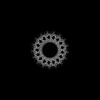 16724MC M: map data used to model this data C: citing same article ( |
|---|---|
| Similar structure data | |
| Experimental dataset #1 | Data reference: 10.6019/EMPIAR-11580 / Data set type: EMPIAR |
-Links
 PDBj
PDBj


- Assembly
| Deposited unit | 
|
|---|---|
| 1 | x 17
|
-Components
-Protein , 6 types, 18 molecules OPQRKLDEFGHIJCBAMN
| #1: Protein | Mass: 19902.945 Da / Num. of mol.: 4 / Source method: isolated from a natural source / Source: (natural) Campylobacter jejuni (Campylobacter) / Strain: 81-176 / References: UniProt: A0A0H3PIF6#2: Protein | | Mass: 91084.125 Da / Num. of mol.: 1 / Source method: isolated from a natural source / Source: (natural) Campylobacter jejuni (Campylobacter) / Strain: 81-176 / References: UniProt: A0A0H3PAU5#3: Protein | | Mass: 93534.625 Da / Num. of mol.: 1 / Source method: isolated from a natural source / Source: (natural) Campylobacter jejuni (Campylobacter) / Strain: 81-176 / References: UniProt: A0A0H3PJ87#4: Protein | Mass: 41455.691 Da / Num. of mol.: 7 / Source method: isolated from a natural source / Source: (natural) Campylobacter jejuni (Campylobacter) / Strain: 81-176 / References: UniProt: A0A1E7NUR8#5: Protein | Mass: 18545.180 Da / Num. of mol.: 3 / Source method: isolated from a natural source / Source: (natural) Campylobacter jejuni (Campylobacter) / Strain: 81-176 / References: UniProt: A0A0H3PCP8#6: Protein | Mass: 27817.662 Da / Num. of mol.: 2 / Source method: isolated from a natural source / Source: (natural) Campylobacter jejuni (Campylobacter) / Strain: 81-176 / References: UniProt: A0A0H3PBX6 |
|---|
-Details
| Has protein modification | N |
|---|
-Experimental details
-Experiment
| Experiment | Method: ELECTRON MICROSCOPY |
|---|---|
| EM experiment | Aggregation state: PARTICLE / 3D reconstruction method: single particle reconstruction |
- Sample preparation
| Component | Name: Periplasmic scaffold structures of the Campylobacter jejuni flagellar motor Type: ORGANELLE OR CELLULAR COMPONENT / Entity ID: all / Source: NATURAL |
|---|---|
| Source (natural) | Organism: Campylobacter jejuni (Campylobacter) / Strain: 81-176 |
| Buffer solution | pH: 7.4 |
| Specimen | Embedding applied: NO / Shadowing applied: NO / Staining applied: NO / Vitrification applied: YES |
| Specimen support | Grid type: Quantifoil R2/2 |
| Vitrification | Cryogen name: ETHANE |
- Electron microscopy imaging
| Experimental equipment |  Model: Titan Krios / Image courtesy: FEI Company | |||||||||||||||
|---|---|---|---|---|---|---|---|---|---|---|---|---|---|---|---|---|
| Microscopy | Model: TFS KRIOS | |||||||||||||||
| Electron gun | Electron source:  FIELD EMISSION GUN / Accelerating voltage: 300 kV / Illumination mode: FLOOD BEAM FIELD EMISSION GUN / Accelerating voltage: 300 kV / Illumination mode: FLOOD BEAM | |||||||||||||||
| Electron lens | Mode: BRIGHT FIELD / Nominal defocus max: 3000 nm / Nominal defocus min: 1500 nm | |||||||||||||||
| Image recording |
| |||||||||||||||
| Image scans |
|
- Processing
| EM software |
| ||||||||||||||||||||||||||||||||||||||||||||||||||||||||||||||||||||||||||||||||||||||||||||||
|---|---|---|---|---|---|---|---|---|---|---|---|---|---|---|---|---|---|---|---|---|---|---|---|---|---|---|---|---|---|---|---|---|---|---|---|---|---|---|---|---|---|---|---|---|---|---|---|---|---|---|---|---|---|---|---|---|---|---|---|---|---|---|---|---|---|---|---|---|---|---|---|---|---|---|---|---|---|---|---|---|---|---|---|---|---|---|---|---|---|---|---|---|---|---|---|
| CTF correction | Type: PHASE FLIPPING AND AMPLITUDE CORRECTION | ||||||||||||||||||||||||||||||||||||||||||||||||||||||||||||||||||||||||||||||||||||||||||||||
| Symmetry | Point symmetry: C17 (17 fold cyclic) | ||||||||||||||||||||||||||||||||||||||||||||||||||||||||||||||||||||||||||||||||||||||||||||||
| 3D reconstruction | Resolution: 7.9 Å / Resolution method: FSC 0.143 CUT-OFF / Num. of particles: 32790 / Symmetry type: POINT | ||||||||||||||||||||||||||||||||||||||||||||||||||||||||||||||||||||||||||||||||||||||||||||||
| Atomic model building | Protocol: FLEXIBLE FIT | ||||||||||||||||||||||||||||||||||||||||||||||||||||||||||||||||||||||||||||||||||||||||||||||
| Atomic model building |
|