Biotechnology and Biological Sciences Research Council (BBSRC)
BB/M011178/1
United Kingdom
Medical Research Council (MRC, United Kingdom)
MR/V000799/1
United Kingdom
National Institutes of Health/National Institute Of Allergy and Infectious Diseases (NIH/NIAID)
R01AI065539
United States
Cancer Research UK
FC001143
United Kingdom
Wellcome Trust
FC001143
United Kingdom
Medical Research Council (MRC, United Kingdom)
FC001143
United Kingdom
Citation
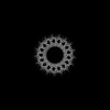
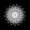
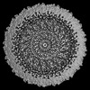
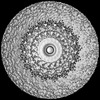
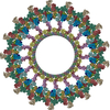
Journal: bioRxiv / Year: 2024 Title: Molecular model of a bacterial flagellar motor reveals a "parts-list" of protein adaptations to increase torque. Authors: Tina Drobnič / Eli J Cohen / Tom Calcraft / Mona Alzheimer / Kathrin Froschauer / Sarah Svensson / William H Hoffmann / Nanki Singh / Sriram G Garg / Louie Henderson / Trishant R Umrekar / ...Authors: Tina Drobnič / Eli J Cohen / Tom Calcraft / Mona Alzheimer / Kathrin Froschauer / Sarah Svensson / William H Hoffmann / Nanki Singh / Sriram G Garg / Louie Henderson / Trishant R Umrekar / Andrea Nans / Deborah Ribardo / Francesco Pedaci / Ashley L Nord / Georg K A Hochberg / David R Hendrixson / Cynthia M Sharma / Peter B Rosenthal / Morgan Beeby / Abstract: One hurdle to understanding how molecular machines work, and how they evolve, is our inability to see their structures . Here we describe a minicell system that enables cryogenic electron microscopy ...One hurdle to understanding how molecular machines work, and how they evolve, is our inability to see their structures . Here we describe a minicell system that enables cryogenic electron microscopy imaging and single particle analysis to investigate the structure of an iconic molecular machine, the bacterial flagellar motor, which spins a helical propeller for propulsion. We determine the structure of the high-torque motor including the subnanometre-resolution structure of the periplasmic scaffold, an adaptation essential to high torque. Our structure enables identification of new proteins, and interpretation with molecular models highlights origins of new components, reveals modifications of the conserved motor core, and explain how these structures both template a wider ring of motor proteins, and buttress the motor during swimming reversals. We also acquire insights into universal principles of flagellar torque generation. This approach is broadly applicable to other membrane-residing bacterial molecular machines complexes.
In the structure databanks used in Yorodumi, some data are registered as the other names, "COVID-19 virus" and "2019-nCoV". Here are the details of the virus and the list of structure data.
Jan 31, 2019. EMDB accession codes are about to change! (news from PDBe EMDB page)
EMDB accession codes are about to change! (news from PDBe EMDB page)
The allocation of 4 digits for EMDB accession codes will soon come to an end. Whilst these codes will remain in use, new EMDB accession codes will include an additional digit and will expand incrementally as the available range of codes is exhausted. The current 4-digit format prefixed with “EMD-” (i.e. EMD-XXXX) will advance to a 5-digit format (i.e. EMD-XXXXX), and so on. It is currently estimated that the 4-digit codes will be depleted around Spring 2019, at which point the 5-digit format will come into force.
The EM Navigator/Yorodumi systems omit the EMD- prefix.
Related info.:Q: What is EMD? / ID/Accession-code notation in Yorodumi/EM Navigator
Yorodumi is a browser for structure data from EMDB, PDB, SASBDB, etc.
This page is also the successor to EM Navigator detail page, and also detail information page/front-end page for Omokage search.
The word "yorodu" (or yorozu) is an old Japanese word meaning "ten thousand". "mi" (miru) is to see.
Related info.:EMDB / PDB / SASBDB / Comparison of 3 databanks / Yorodumi Search / Aug 31, 2016. New EM Navigator & Yorodumi / Yorodumi Papers / Jmol/JSmol / Function and homology information / Changes in new EM Navigator and Yorodumi
 Movie
Movie Controller


 Open data
 Basic information
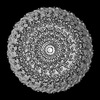
 Map data
 Sample
 Keywords
 Campylobacter jejuni (Campylobacter)
 Authors
 United Kingdom,
  United States, 6 items
  Citation

 Structure visualization
 Downloads & links
 EMDB map data format
 emd_17416.png
 http://ftp.pdbj.org/pub/emdb/structures/EMD-17416
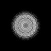
 Z (Sec.)
 Y (Row.)
 X (Col.)


 Sample components
 Processing
 Electron microscopy
 FIELD EMISSION GUN

