[English] 日本語
Yorodumi
- PDB-9hdb: Sla1 SH3_3 domain (residues 355-414) -

+
Open data


ID or keywords:

Loading...

-
Basic information

Entry
Database: PDB / ID: 9hdb
TitleSla1 SH3_3 domain (residues 355-414)
ComponentsActin cytoskeleton-regulatory complex protein SLA1
KeywordsENDOCYTOSIS / SH3 domain / Membrane trafficking / protein-protein interaction
Function / homology
Function and homology information


actin cytoskeleton-regulatory complex / SLAC complex / cargo adaptor activity / RND2 GTPase cycle / RHOQ GTPase cycle / actin cortical patch assembly / regulation of Arp2/3 complex-mediated actin nucleation / negative regulation of Arp2/3 complex-mediated actin nucleation / actin cortical patch / regulation of actin filament polymerization ...actin cytoskeleton-regulatory complex / SLAC complex / cargo adaptor activity / RND2 GTPase cycle / RHOQ GTPase cycle / actin cortical patch assembly / regulation of Arp2/3 complex-mediated actin nucleation / negative regulation of Arp2/3 complex-mediated actin nucleation / actin cortical patch / regulation of actin filament polymerization / cellular bud neck / mating projection tip / ubiquitin binding / cell wall organization / endocytosis / actin binding / cell cortex / endosome membrane / identical protein binding / nucleus / plasma membrane / cytoplasm
Similarity search - Function
SLA1 homology domain 1, SHD1 / Sla1, first SH3 domain / Sla1, third SH3 domain / SLA1 homology domain 1, SHD1 / SLA1-like, PH domain-like / Variant SH3 domain / Sterile alpha motif/pointed domain superfamily / SH3 domain / Src homology 3 domains / SH3-like domain superfamily ...SLA1 homology domain 1, SHD1 / Sla1, first SH3 domain / Sla1, third SH3 domain / SLA1 homology domain 1, SHD1 / SLA1-like, PH domain-like / Variant SH3 domain / Sterile alpha motif/pointed domain superfamily / SH3 domain / Src homology 3 domains / SH3-like domain superfamily / Src homology 3 (SH3) domain profile. / SH3 domain
Similarity search - Domain/homology
Actin cytoskeleton-regulatory complex protein SLA1
Similarity search - Component
Biological speciesSaccharomyces cerevisiae (brewer's yeast)
MethodX-RAY DIFFRACTION / SYNCHROTRON / MOLECULAR REPLACEMENT / Resolution: 1.491 Å
AuthorsDraper-Barr, G. / Defelipe, L.A. / Ruiz-Carillo, D. / Garcia-Alai, M.M.
Funding support Germany, 1items
OrganizationGrant numberCountry
EIPOD fellowship under Marie Sklodowska-Curie Actions COFUND664726 Germany
Citation
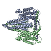
Journal: Structure / Year: 2025
Title: Sla2 is a core interaction hub for clathrin light chain and the Pan1/End3/Sla1 complex.
Authors: George Draper-Barr / Lucas A Defelipe / David Ruiz-Carrillo / Emil Gustavsson / Meytal Landau / Maria García-Alai /
Abstract: The interaction network of Sla2, a vital endocytic mid-coat adaptor protein, undergoes constant rearrangement. Sla2 serves as a scaffold linking the membrane to the actin cytoskeleton, with its role ...The interaction network of Sla2, a vital endocytic mid-coat adaptor protein, undergoes constant rearrangement. Sla2 serves as a scaffold linking the membrane to the actin cytoskeleton, with its role modulated by the clathrin light chain (CLC), which inhibits Sla2's function under certain conditions. We show that Sla2 has two independent binding sites for CLC: one previously described in homologs of fungi (Sla2) and metazoa (Hip1R), and a second found only in Fungi. We present the structural model of the Sla2 actin-binding domains in the context of regulatory structural domains by cryoelectron microscopy. We provide an interaction map of Sla2 and the regulatory proteins Sla1 and Pan1, predicted by AI modeling and confirmed by molecular biophysics techniques. Pan1 may compete with CLC for the conserved Sla2-binding site. These results enhance the mapping of crucial interactions at endocytic checkpoints and highlight the divergence between Metazoa and Fungi in this vital process.
History
DepositionNov 12, 2024Deposition site: PDBE / Processing site: PDBE
Revision 1.0May 21, 2025Provider: repository / Type: Initial release

-
Structure visualization

Structure viewerMolecule:
MolmilJmol/JSmol

Downloads & links

-
Assembly

Deposited unit
B: Actin cytoskeleton-regulatory complex protein SLA1
A: Actin cytoskeleton-regulatory complex protein SLA1


Theoretical massNumber of molelcules
Total (without water)14,5252
Polymers14,5252
Non-polymers00
Water2,000111
1
B: Actin cytoskeleton-regulatory complex protein SLA1


Theoretical massNumber of molelcules
Total (without water)7,2621
Polymers7,2621
Non-polymers00
Water181
TypeNameSymmetry operationNumber
identity operation1_555x,y,z1
2
A: Actin cytoskeleton-regulatory complex protein SLA1


Theoretical massNumber of molelcules
Total (without water)7,2621
Polymers7,2621
Non-polymers00
Water181
TypeNameSymmetry operationNumber
identity operation1_555x,y,z1
Unit cell
Length a, b, c (Å)38.533, 50.423, 51.967
Angle α, β, γ (deg.)90, 90, 90
Int Tables number19
Space group name H-MP212121
Noncrystallographic symmetry (NCS)NCS domain:
IDEns-IDDetails
11B
21A

NCS domain segments:

Component-ID: 1 / Ens-ID: 1 / Beg auth comp-ID: LYS / Beg label comp-ID: LYS / End auth comp-ID: VAL / End label comp-ID: VAL / Auth seq-ID: 6 - 63 / Label seq-ID: 6 - 63

Dom-IDAuth asym-IDLabel asym-ID
1BA
2AB

NCS ensembles : (Details: Local NCS retraints between domains: 1 2)

-
Components

#1: Protein Actin cytoskeleton-regulatory complex protein SLA1


Mass: 7262.365 Da / Num. of mol.: 2
Source method: isolated from a genetically manipulated source
Details: residues 355-414 of Sla1. Purified from an N-terminal GST fusion construct leaving the GAMA sequence prior to the domain of interest after TEV cleavage.
Source: (gene. exp.) Saccharomyces cerevisiae (brewer's yeast)
Gene: SLA1, YBL007C, YBL0321 / Plasmid: pETM30
Details (production host): 6xHis-GST-'TEV cleavage site'-POI
Production host: Escherichia coli (E. coli) / References: UniProt: P32790
#2: Water ChemComp-HOH / water


Mass: 18.015 Da / Num. of mol.: 111 / Source method: isolated from a natural source / Formula: H2O
Has protein modificationN

-
Experimental details

-
Experiment

ExperimentMethod: X-RAY DIFFRACTION / Number of used crystals: 1

-
Sample preparation

CrystalDensity Matthews: 1.74 Å3/Da / Density % sol: 29.22 %
Crystal growTemperature: 292 K / Method: vapor diffusion, sitting drop / pH: 7.5
Details: 0.1M HEPES pH 7.5 20% PEG 10000 1mM peptide (PVSTPARTPARTPTP) dissolved in 0.03M HEPES pH 8, 0.15M NaCl, 0.5mM TCEP

-
Data collection

DiffractionMean temperature: 100 K / Serial crystal experiment: N
Diffraction sourceSource: SYNCHROTRON / Site: PETRA III, EMBL c/o DESY / Beamline: P13 (MX1) / Wavelength: 0.9762 Å
DetectorType: DECTRIS PILATUS 6M-F / Detector: PIXEL / Date: Oct 22, 2024
RadiationMonochromator: Si(111) / Protocol: SINGLE WAVELENGTH / Monochromatic (M) / Laue (L): M / Scattering type: x-ray
Radiation wavelengthWavelength: 0.9762 Å / Relative weight: 1
ReflectionResolution: 1.49→51.97 Å / Num. obs: 17131 / % possible obs: 100 % / Redundancy: 10 % / CC1/2: 0.998 / Rmerge(I) obs: 0.078 / Rpim(I) all: 0.037 / Rrim(I) all: 0.087 / Χ2: 0.98 / Net I/σ(I): 15.6
Reflection shell

Χ2: 0.98 / Diffraction-ID: 1

Resolution (Å)Redundancy (%)Rmerge(I) obsMean I/σ(I) obsNum. unique obsCC1/2Rpim(I) allRrim(I) all% possible all
8.16-51.977.20.07124.21390.9890.0360.0899.7
1.49-1.529.30.4224.88200.9430.2110.47399.9

-
Processing

Software
NameVersionClassification
REFMAC5.8.0430 (refmacat 0.4.88)refinement
Aimlessdata scaling
autoPROCdata reduction
PHENIXmodel building
Cootmodel building
MOLREPphasing
RefinementMethod to determine structure: MOLECULAR REPLACEMENT / Resolution: 1.491→36.214 Å / Cor.coef. Fo:Fc: 0.971 / Cor.coef. Fo:Fc free: 0.961 / SU B: 2.543 / SU ML: 0.044 / Cross valid method: FREE R-VALUE / ESU R: 0.09 / ESU R Free: 0.075
Details: Hydrogens have been added in their riding positions
RfactorNum. reflection% reflectionSelection details
Rfree0.1895 848 4.964 %RANDOM
Rwork0.143 16235 --
all0.145 ---
obs-17083 99.947 %-
Solvent computationIon probe radii: 0.8 Å / Shrinkage radii: 0.8 Å / VDW probe radii: 1.2 Å / Solvent model: MASK BULK SOLVENT
Displacement parametersBiso mean: 18.897 Å2
Baniso -1Baniso -2Baniso -3
1--0.915 Å20 Å20 Å2
2--0.226 Å2-0 Å2
3---0.689 Å2
Refinement stepCycle: LAST / Resolution: 1.491→36.214 Å
ProteinNucleic acidLigandSolventTotal
Num. atoms982 0 0 111 1093
Refine LS restraints
Refine-IDTypeDev idealDev ideal targetNumber
X-RAY DIFFRACTIONr_bond_refined_d0.0150.0121007
X-RAY DIFFRACTIONr_bond_other_d0.0010.016967
X-RAY DIFFRACTIONr_angle_refined_deg1.9651.8091354
X-RAY DIFFRACTIONr_angle_other_deg0.6641.8092244
X-RAY DIFFRACTIONr_dihedral_angle_1_deg6.1535123
X-RAY DIFFRACTIONr_dihedral_angle_2_deg26.04854
X-RAY DIFFRACTIONr_dihedral_angle_3_deg13.14510195
X-RAY DIFFRACTIONr_dihedral_angle_6_deg16.8851044
X-RAY DIFFRACTIONr_chiral_restr0.1060.2143
X-RAY DIFFRACTIONr_gen_planes_refined0.010.021169
X-RAY DIFFRACTIONr_gen_planes_other0.0010.02215
X-RAY DIFFRACTIONr_nbd_refined0.1960.2125
X-RAY DIFFRACTIONr_symmetry_nbd_other0.1940.2854
X-RAY DIFFRACTIONr_nbtor_refined0.180.2470
X-RAY DIFFRACTIONr_symmetry_nbtor_other0.0890.2561
X-RAY DIFFRACTIONr_xyhbond_nbd_refined0.1560.262
X-RAY DIFFRACTIONr_symmetry_nbd_refined0.1570.218
X-RAY DIFFRACTIONr_nbd_other0.1550.264
X-RAY DIFFRACTIONr_symmetry_xyhbond_nbd_refined0.190.226
X-RAY DIFFRACTIONr_mcbond_it4.41.092495
X-RAY DIFFRACTIONr_mcbond_other4.3751.091495
X-RAY DIFFRACTIONr_mcangle_it5.9921.963617
X-RAY DIFFRACTIONr_mcangle_other5.9951.964618
X-RAY DIFFRACTIONr_scbond_it6.2141.322512
X-RAY DIFFRACTIONr_scbond_other6.2081.321513
X-RAY DIFFRACTIONr_scangle_it8.5452.334737
X-RAY DIFFRACTIONr_scangle_other8.542.332738
X-RAY DIFFRACTIONr_lrange_it11.56616.5414028
X-RAY DIFFRACTIONr_lrange_other11.09715.693956
X-RAY DIFFRACTIONr_rigid_bond_restr5.64431974
X-RAY DIFFRACTIONr_ncsr_local_group_10.1470.051719
Refine LS restraints NCS
Ens-IDDom-IDAuth asym-IDRefine-IDTypeRms dev position (Å)Weight position
11BX-RAY DIFFRACTIONLocal ncs0.14680.05008
12AX-RAY DIFFRACTIONLocal ncs0.14680.05008
LS refinement shell

Refine-ID: X-RAY DIFFRACTION / Total num. of bins used: 20

Resolution (Å)Rfactor RfreeNum. reflection RfreeRfactor RworkNum. reflection RworkRfactor allNum. reflection allFsc freeFsc work% reflection obs (%)WRfactor Rwork
1.491-1.5290.211520.12911800.13212330.9790.9999.91890.119
1.529-1.5710.235830.10611160.11411990.9650.9921000.099
1.571-1.6170.168610.09711280.10111890.9830.9941000.089
1.617-1.6660.179670.10610560.1111250.9770.99399.82220.098
1.666-1.7210.216480.11610720.1211200.9720.9921000.107
1.721-1.7810.2500.11810160.12210670.9780.99199.90630.111
1.781-1.8480.212360.11910100.12210470.9690.99199.90450.115
1.848-1.9230.163370.1259720.12610090.9820.991000.122
1.923-2.0090.203420.129220.1249640.9740.9911000.121
2.009-2.1060.177630.1218560.1259210.9830.99199.78280.126
2.106-2.220.195530.1248220.1288750.9820.9911000.129
2.22-2.3540.233340.1358080.1398420.9750.9891000.143
2.354-2.5160.188420.1427420.1457840.980.9871000.152
2.516-2.7160.216340.1487060.1517400.9720.9861000.161
2.716-2.9740.177450.1466400.1486850.9820.9871000.166
2.974-3.3220.145310.1435980.1436300.9880.98699.84130.164
3.322-3.830.195220.1515350.1535580.9820.98699.82080.179
3.83-4.6780.144210.1284570.1294780.9890.991000.167
4.678-6.5610.198210.1993670.1993880.9730.9771000.241
6.561-36.2140.3760.322320.3212380.9590.9381000.415
Refinement TLS params.

Method: refined / Refine-ID: X-RAY DIFFRACTION

IDL112)L122)L132)L222)L232)L332)S11 (Å °)S12 (Å °)S13 (Å °)S21 (Å °)S22 (Å °)S23 (Å °)S31 (Å °)S32 (Å °)S33 (Å °)T112)T122)T132)T222)T232)T332)Origin x (Å)Origin y (Å)Origin z (Å)
11.93280.35270.07372.66270.07522.32780.0181-0.06790.02550.03030.0016-0.07610.05370.0542-0.01970.00840.00120.00130.0188-0.00560.0049-9.7501-5.2405-9.1819
23.36240.2839-0.36912.61160.56133.36460.0186-0.01170.18370.0057-0.0290.0646-0.1388-0.07310.01040.00640.00190.00010.0045-0.00120.01118.8075-6.9381-16.864
Refinement TLS group
IDRefine-IDRefine TLS-IDSelectionAuth asym-IDAuth seq-ID
1X-RAY DIFFRACTION1ALLBp1 - 64
2X-RAY DIFFRACTION2ALLAp6 - 64

+
About Yorodumi

-
News

-
Feb 9, 2022. New format data for meta-information of EMDB entries

New format data for meta-information of EMDB entries

  • Version 3 of the EMDB header file is now the official format.
  • The previous official version 1.9 will be removed from the archive.

Related info.:EMDB header

External links:wwPDB to switch to version 3 of the EMDB data model

-
Aug 12, 2020. Covid-19 info

Covid-19 info

URL: https://pdbj.org/emnavi/covid19.php

New page: Covid-19 featured information page in EM Navigator.

Related info.:Covid-19 info / Mar 5, 2020. Novel coronavirus structure data

+
Mar 5, 2020. Novel coronavirus structure data

Novel coronavirus structure data

Related info.:Yorodumi Speices / Aug 12, 2020. Covid-19 info

External links:COVID-19 featured content - PDBj / Molecule of the Month (242):Coronavirus Proteases

+
Jan 31, 2019. EMDB accession codes are about to change! (news from PDBe EMDB page)

EMDB accession codes are about to change! (news from PDBe EMDB page)

  • The allocation of 4 digits for EMDB accession codes will soon come to an end. Whilst these codes will remain in use, new EMDB accession codes will include an additional digit and will expand incrementally as the available range of codes is exhausted. The current 4-digit format prefixed with “EMD-” (i.e. EMD-XXXX) will advance to a 5-digit format (i.e. EMD-XXXXX), and so on. It is currently estimated that the 4-digit codes will be depleted around Spring 2019, at which point the 5-digit format will come into force.
  • The EM Navigator/Yorodumi systems omit the EMD- prefix.

Related info.:Q: What is EMD? / ID/Accession-code notation in Yorodumi/EM Navigator

External links:EMDB Accession Codes are Changing Soon! / Contact to PDBj

+
Jul 12, 2017. Major update of PDB

Major update of PDB

  • wwPDB released updated PDB data conforming to the new PDBx/mmCIF dictionary.
  • This is a major update changing the version number from 4 to 5, and with Remediation, in which all the entries are updated.
  • In this update, many items about electron microscopy experimental information are reorganized (e.g. em_software).
  • Now, EM Navigator and Yorodumi are based on the updated data.

External links:wwPDB Remediation / Enriched Model Files Conforming to OneDep Data Standards Now Available in the PDB FTP Archive

-
Yorodumi

Thousand views of thousand structures

  • Yorodumi is a browser for structure data from EMDB, PDB, SASBDB, etc.
  • This page is also the successor to EM Navigator detail page, and also detail information page/front-end page for Omokage search.
  • The word "yorodu" (or yorozu) is an old Japanese word meaning "ten thousand". "mi" (miru) is to see.

Related info.:EMDB / PDB / SASBDB / Comparison of 3 databanks / Yorodumi Search / Aug 31, 2016. New EM Navigator & Yorodumi / Yorodumi Papers / Jmol/JSmol / Function and homology information / Changes in new EM Navigator and Yorodumi

Read more