 Movie
Movie Controller

[English] 日本語
 Yorodumi
- EMDB-52061: Sla2 C-terminal region (Residues 560-968) (REND and THATCH domains) -
+ Open data

- Basic information
| Entry | 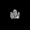 | |||||||||
|---|---|---|---|---|---|---|---|---|---|---|
| Title | Sla2 C-terminal region (Residues 560-968) (REND and THATCH domains) | |||||||||
 Map data Map data | ||||||||||
 Sample Sample |
| |||||||||
 Keywords Keywords | Actin binding / Endocytosis / membrane trafficking | |||||||||
| Function / homology |  Function and homology information Function and homology informationactin cortical patch assembly / clathrin light chain binding / incipient cellular bud site / actin cortical patch / cellular bud tip / negative regulation of Arp2/3 complex-mediated actin nucleation / clathrin coat assembly / clathrin-cargo adaptor activity / cellular bud neck / mating projection tip ...actin cortical patch assembly / clathrin light chain binding / incipient cellular bud site / actin cortical patch / cellular bud tip / negative regulation of Arp2/3 complex-mediated actin nucleation / clathrin coat assembly / clathrin-cargo adaptor activity / cellular bud neck / mating projection tip / phosphatidylinositol-3,4-bisphosphate binding / phosphatidylinositol-3,5-bisphosphate binding / clathrin-coated vesicle / cortical actin cytoskeleton / actin filament organization / endocytosis / actin filament binding / plasma membrane Similarity search - Function | |||||||||
| Biological species |  | |||||||||
| Method | single particle reconstruction / cryo EM / Resolution: 3.62 Å | |||||||||
 Authors Authors | Draper-Barr G / Gustavsson E / Landau M / Garcia-Alai MM | |||||||||
| Funding support |  Germany, 1 items Germany, 1 items
| |||||||||
 Citation Citation | Journal: Structure / Year: 2025 Title: Sla2 is a core interaction hub for clathrin light chain and the Pan1/End3/Sla1 complex. Authors: George Draper-Barr / Lucas A Defelipe / David Ruiz-Carrillo / Emil Gustavsson / Meytal Landau / Maria García-Alai /  Abstract: The interaction network of Sla2, a vital endocytic mid-coat adaptor protein, undergoes constant rearrangement. Sla2 serves as a scaffold linking the membrane to the actin cytoskeleton, with its role ...The interaction network of Sla2, a vital endocytic mid-coat adaptor protein, undergoes constant rearrangement. Sla2 serves as a scaffold linking the membrane to the actin cytoskeleton, with its role modulated by the clathrin light chain (CLC), which inhibits Sla2's function under certain conditions. We show that Sla2 has two independent binding sites for CLC: one previously described in homologs of fungi (Sla2) and metazoa (Hip1R), and a second found only in Fungi. We present the structural model of the Sla2 actin-binding domains in the context of regulatory structural domains by cryoelectron microscopy. We provide an interaction map of Sla2 and the regulatory proteins Sla1 and Pan1, predicted by AI modeling and confirmed by molecular biophysics techniques. Pan1 may compete with CLC for the conserved Sla2-binding site. These results enhance the mapping of crucial interactions at endocytic checkpoints and highlight the divergence between Metazoa and Fungi in this vital process. | |||||||||
| History |
|
- Structure visualization
| Supplemental images |
|---|

- Downloads & links
-EMDB archive
| Map data | emd_52061.map.gz | 9.3 MB | EMDB map data format | |
|---|---|---|---|---|
| Header (meta data) | emd-52061-v30.xmlemd-52061.xml | 20.1 KB 20.1 KB | Display Display | EMDB header |
| FSC (resolution estimation) | emd_52061_fsc.xml | 5.6 KB | Display | FSC data file |
| Images |  emd_52061.png emd_52061.png | 68.9 KB | ||
| Masks | emd_52061_msk_1.map | 18.7 MB | Mask map | |
| Filedesc metadata | emd-52061.cif.gz | 6.8 KB | ||
| Others | emd_52061_half_map_1.map.gzemd_52061_half_map_2.map.gz | 17.4 MB 17.4 MB | ||
| Archive directory |  http://ftp.pdbj.org/pub/emdb/structures/EMD-52061ftp://ftp.pdbj.org/pub/emdb/structures/EMD-52061 http://ftp.pdbj.org/pub/emdb/structures/EMD-52061ftp://ftp.pdbj.org/pub/emdb/structures/EMD-52061 | HTTPS FTP |
-Related structure data
| Related structure data | 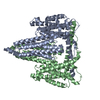 9hddMC  9hdbC M: atomic model generated by this map C: citing same article ( |
|---|---|
| Similar structure data |
-Links
| EMDB pages | EMDB (EBI/PDBe) / EMDataResource |
|---|---|
| Related items in Molecule of the Month |


-Map
| File | Download / File: emd_52061.map.gz / Format: CCP4 / Size: 18.7 MB / Type: IMAGE STORED AS FLOATING POINT NUMBER (4 BYTES) | ||||||||||||||||||||||||||||||||||||
|---|---|---|---|---|---|---|---|---|---|---|---|---|---|---|---|---|---|---|---|---|---|---|---|---|---|---|---|---|---|---|---|---|---|---|---|---|---|
| Projections & slices | Image control
Images are generated by Spider. | ||||||||||||||||||||||||||||||||||||
| Voxel size | X=Y=Z: 1.36 Å | ||||||||||||||||||||||||||||||||||||
| Density |
| ||||||||||||||||||||||||||||||||||||
| Symmetry | Space group: 1 | ||||||||||||||||||||||||||||||||||||
| Details | EMDB XML:
|
 Z (Sec.)
 Y (Row.)
 X (Col.)


-Supplemental data
-Mask #1
| File | emd_52061_msk_1.map | ||||||||||||
|---|---|---|---|---|---|---|---|---|---|---|---|---|---|
| Projections & Slices |
| ||||||||||||
| Density Histograms |


-Half map: #2
| File | emd_52061_half_map_1.map | ||||||||||||
|---|---|---|---|---|---|---|---|---|---|---|---|---|---|
| Projections & Slices |
| ||||||||||||
| Density Histograms |


-Half map: #1
| File | emd_52061_half_map_2.map | ||||||||||||
|---|---|---|---|---|---|---|---|---|---|---|---|---|---|
| Projections & Slices |
| ||||||||||||
| Density Histograms |


- Sample components
-Entire : ScSla2 residues 351-968
| Entire | Name: ScSla2 residues 351-968 |
|---|---|
| Components |
|
-Supramolecule #1: ScSla2 residues 351-968
| Supramolecule | Name: ScSla2 residues 351-968 / type: complex / ID: 1 / Parent: 0 / Macromolecule list: all Details: ScSla2:351-968 forms a dimer through the coiled-coil and REND domain between residues 351-735. |
|---|---|
| Source (natural) | Organism: |
| Molecular weight | Theoretical: 140 KDa |
-Macromolecule #1: Protein SLA2
| Macromolecule | Name: Protein SLA2 / type: protein_or_peptide / ID: 1 Details: Complete expression construct was residues 351-968 of ScSla2. Only residues 560-968 were able to be modelled into the electron density due to the inherent flexibility of the residues 351-559 ...Details: Complete expression construct was residues 351-968 of ScSla2. Only residues 560-968 were able to be modelled into the electron density due to the inherent flexibility of the residues 351-559 that form the coiled-coil. Number of copies: 2 / Enantiomer: LEVO |
|---|---|
| Source (natural) | Organism: |
| Molecular weight | Theoretical: 69.678672 KDa |
| Recombinant expression | Organism:  |
| Sequence | String: ATAQMQPDFW ANQQAQFANE QNRLEQERVQ QLQQQQAQQE LFQQQLQKAQ QDMMNMQLQQ QNQHQNDLIA LTNQYEKDQA LLQQYDQRV QQLESEITTM DSTASKQLAN KDEQLTALQD QLDVWERKYE SLAKLYSQLR QEHLNLLPRF KKLQLKVNSA Q ESIQKKEQ ...String: ATAQMQPDFW ANQQAQFANE QNRLEQERVQ QLQQQQAQQE LFQQQLQKAQ QDMMNMQLQQ QNQHQNDLIA LTNQYEKDQA LLQQYDQRV QQLESEITTM DSTASKQLAN KDEQLTALQD QLDVWERKYE SLAKLYSQLR QEHLNLLPRF KKLQLKVNSA Q ESIQKKEQ LEHKLKQKDL QMAELVKDRD RARLELERSI NNAEADSAAA TAAAETMTQD KMNPILDAIL ESGINTIQES VY NLDSPLS WSGPLTPPTF LLSLLESTSE NATEFATSFN NLIVDGLAHG DQTEVIHCVS DFSTSMATLV TNSKAYAVTT LPQ EQSDQI LTLVKRCARE AQYFFEDLMS ENLNQVGDEE KTDIVINANV DMQEKLQELS LAIEPLLNIQ SVKSNKETNP HSEL VATAD KIVKSSEHLR VDVPKPLLSL ALMIIDAVVA LVKAAIQCQN EIATTTSIPL NQFYLKNSRW TEGLISAAKA VAGAT NVLI TTASKLITSE DNENTSPEQF IVASKEVAAS TIQLVAASRV KTSIHSKAQD KLEHCSKDVT DACRSLGNHV MGMIED DHS TSQQQQPLDF TSEHTLKTAE MEQQVEILKL EQSLSNARKR LGEIRRHAYY NQDDD UniProtKB: Protein SLA2 |
-Experimental details
-Structure determination
| Method | cryo EM |
|---|---|
 Processing Processing | single particle reconstruction |
| Aggregation state | particle |
-Sample preparation
| Concentration | 2 mg/mL | ||||||||||||
|---|---|---|---|---|---|---|---|---|---|---|---|---|---|
| Buffer | pH: 8 Component:
Details: 0.03 M HEPES pH 8 0.15 M NaCl 0.5 mM TCEP 0.1 um filtered buffer and degassed for one hour at room temperature | ||||||||||||
| Grid | Model: Quantifoil R2/2 / Material: GOLD / Mesh: 300 / Support film - Material: CARBON / Pretreatment - Type: GLOW DISCHARGE / Pretreatment - Time: 60 sec. | ||||||||||||
| Vitrification | Cryogen name: ETHANE-PROPANE / Chamber humidity: 95 % / Chamber temperature: 279 K / Instrument: FEI VITROBOT MARK IV | ||||||||||||
| Details | monodisperse dimers of the ScSla2:351-968 construct |
- Electron microscopy
| Microscope | TFS KRIOS |
|---|---|
| Image recording | Film or detector model: GATAN K3 (6k x 4k) / Number grids imaged: 1 / Average electron dose: 45.0 e/Å2 |
| Electron beam | Acceleration voltage: 300 kV / Electron source:  FIELD EMISSION GUN FIELD EMISSION GUN |
| Electron optics | C2 aperture diameter: 70.0 µm / Illumination mode: SPOT SCAN / Imaging mode: BRIGHT FIELD / Cs: 2.7 mm / Nominal defocus max: 2.0 µm / Nominal defocus min: 0.5 µm / Nominal magnification: 120000 |
| Sample stage | Specimen holder model: FEI TITAN KRIOS AUTOGRID HOLDER / Cooling holder cryogen: NITROGEN |
| Experimental equipment |  Model: Titan Krios / Image courtesy: FEI Company |
+Image processing

-Atomic model buiding 1
| Initial model | PDB ID: Chain - Residue range: 560-968 / Chain - Source name: AlphaFold / Chain - Initial model type: in silico model / Details: two chains of this model |
|---|---|
| Refinement | Space: REAL / Protocol: FLEXIBLE FIT / Target criteria: CC |
| Output model | PDB-9hdd: |

